Abstract
Fractures of the bones that make the wrist joint together with injury to the ligaments and joint capsules are frequent traumas. It can cause besides limited movement also the pathological mobility. These mild injuries often do not provide the degree of recognizable symptoms and signs. They are diagnosed by X-ray imaging, stress images. Before arthrography was an important method, but nowadays arthroscopy has the advantage. Fresh bone and ligament injuries can be and should be repaired in the early posttraumatic period. Unrecognized and undiagnosed injuries are leading to instability of the wrist, to motion abnormalities or impingement overload syndrome. In the treatment of instability important place have reconstruction of the ligaments and arthrodesis of the wrist.
Keywords: wrist, anatomy, diagnosis of instability, treatment.
1. INTRODUCTION
Instability of the wrist was first described by Gilford et al in 1943. They first wrote about the connection between the proximal and distal row of carpal bones and pointed to consequential instability of the system under the influence of compressive forces. Later, Fisk in 1970 stressed relationship between the carpal instability and scaphoid bone fracture. Linsheid and associates (1972) and Dobyns et al (1975) have written important articles which have defined the instability of the wrist, described the X-ray findings, and offer the draft classification and the first therapeutic approach. Since then, things have become more complicated, mainly because lack of consensual terminology. The International Wrist Investigators Workshop Nomenclature Committee was established to define the terms used to describe carpal instability, but so far no single system is not harmonized. Larson and colleagues in 1995 made system with six categories, based on chronicity, constancy, etiology, location, direction and mechanism of instability, with the aim to standardize the diagnosis, treatment and uniformity in publishing results (1).
1.1. WRIST ANATOMY
Seven carpal bones (excluding pisiform bone which is sesamoid bone) are arranged in two rows. Proximal row is made of scaphoid, lunate and triquetrum bone. The distal row consists trapezium, trapezoid, capipate and hamate bone, and forms a stable surface on which rely metacarpal bones. Movement between the bones of the distal row is very limited. Scaphoid bone is movable connection between the proximal and distal carpal bones rows. When the movement of the wrist from ulnar to radial deviation, the scaphoid bone is moved in volar direction, to avoid the radius styloid process (this movement is used when performing Kirk-Watson test).
Talesnikov (1985) modification of the Navaro concept of the wrist with longitudinal columns is another way to analyze the same concept. In practice, the concept of columns and rows helps in understanding the carpal mechanism.
Tendons are not attached directly to the wrist. The stability of the joint depends on the shape of bones, ligaments, intrinsic (between carpal bones) and extrinsic ligaments (the volar and dorsal sides of the carpus). Instability occurs when damage occurs to any of these elements.
Carpal intrinsic ligaments between the bones are short. Their break can occur in case of twisting (e.g., compressive forces acting on the outstretched wrist or on a motorcycle accident). Extrinsic ligaments are by far the stronger on the palmar side of the wrist which form two strong “V” shapes. Radioscafolunate ligament is considered to be the stabilizer of proximal scaphoid bone. Intrinsic ligaments are covered by synovia and therefore it is not easy to see them during the surgery (1).
1.2. FUNCTIONAL ANATOMY
Smooth function and joint stability provides healthy, undamaged by trauma or pathologic process joint. Trauma or pathologic process are leading to injuries and damage to bones, ligaments and joint capsules. Articulation is lost, and later are developed the degenerative process in the bones of the wrist in terms of arthrosis. The result is abnormal mobility and instability.
1.3. WRIST INSTABILITY
The first explanation of the term is connected with the name Fisk and Linscheid (2).
“They explained Post-traumatic instability of the wrist with the clinical signs and X-ray changes. Further insights provided clinical research and diagnostic moments (arthrography, arthroscopy, CT)”.
Important factors:
The connection and interaction–the wrist with the first row of bones – joints with second row of wrist bones.
Lunate, scaphoid, and the remaining metacarpal bones of the first row
One group of authors prefers the role of scaphoid bone
Ulnar ligaments and pisiform bone
Radial ligaments and scaphoid bone (one of the most important factor)
Inter-bone ligaments
In the system of stability also participates extraarticular systems–retinaculom extensorum and retinaculom flexorum.
The etiology of instability
Damage to the articular capsule
Ligaments lesions
Scaphoid bone fracture
Directly affects the stability of adjacent bone axis lunate and capitate bone. Lunate bone moves dorsally and capitate bone proximally. In the later course of develops malation of the lunate and shortening. Leading to deformation and wrist movement.
1.4. CLASSIFICITION
Dobyns et al. (1975) described four types of carpal instability: dorsal flexion, palmar flexion, ulnar translocation and dorsal subluxation. Instability can occur as a result of ligaments tearing, or what are rarer, carpal bones fractures (1).
DISI (dorsal flexed intercalate segment instability)
Dorsal flexion instability is the most common type of carpal instability, and usually occurs in case of fall on prone, dorsal flexed wrist. It is manifested by a wide Scafo-lunate gap that is larger than 5 mm. When broken scafolunate ligament is tearing, scaphoid bone crosses in flexion. This is best seen on lateral views, where scafolunate angle greater than 47° (scaphoid bone protrudes more to palmar side). At the antero-posterior X-ray flexed scaphoid bone is shown as a “ring” when the palmar tuberosity is placed more than normal. Intercalate segment refers to the axis lunate, relationship between the radius and carpus, a concept that was first made by Gilford (1943). On lateral views, lunate bone protrudes dorsally as inverted tea cup compared to the radius and flexed scaphoid bone. This is typical of a DISI deformity (1).
If not treated, this ruptured intercarpal ligament becomes deranged carpal mechanism. The cavity between the scaphoid and lunate bone spreads and capitatutum axis begins to descend between the two, with subsequent osteoarthritis, which is known as advanced scapho-lunate collapse or SLAC wrist (Scapho-Lunate Advanced Collapse) (1).
VISI (volar or palmar flexed intercalate segment instability). It is much rarer and occurs at the ulnar side of the lunate bone, at the joint between the lunate and triquetrum bone. The cavity between the lunate and triquetrum bone is not always seen, and the clinical picture is often mixed with injuries in the middle carpus, the joint between the triquetrum and hamate bone. On lateral radiograms, lunate bone is oriented as palmar poured tea cup (1).
2. CLINICAL PRESENTATION
2.1. Anamnesis
It is important to know the patient’s age and occupation. Typically, the data present in the anamnesis are significant to injuries to the wrist, usually in a position dorsal flexion with joint swelling. However, the clinical picture often occurs with delay, with a vague pain in the joints and weakened grip. Typically there is no bone fractures, although carpal instability is described as in in case of frakturae radii loco tipico (Colles fracture) (1).
2.2. Symptoms
There is pain in the wrist, localized mainly in the area of the scaphoid bone, edema in that region, which is expressed during the first few days after the trauma and feeling of instability and uncertainty within the wrist (3).
2.3. Physical examination
The area that id softest should be first determined and it is best to be avoided in the first examination in order to gain the trust of the patient. You should apply the wellknown Apley approach “look, feel, move”. Full carpus should be palpated, starting from the radial and volar part of the wrist, where the scaphoid tuberosity is palpated on the basis of the tendon of M. flexor carpi radialis (1).
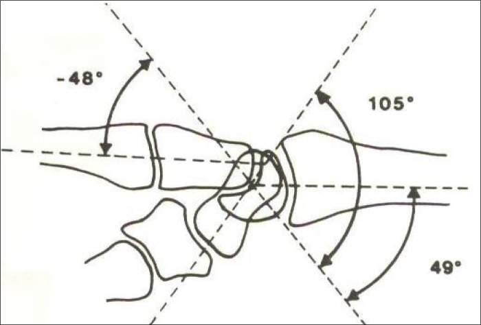
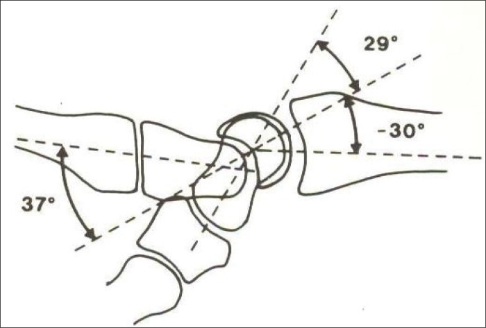
Each bone of the wrist, like any ligament that is palpable, it should be palpable. Provoking maneuvers that act on the intrinsic and extrinsic ligaments can also be performed. (4) (Figure 1 and 2).
Figure 1.
Schematic view of the dorsal angle of wrist instability “DISI”, Source: Buck and Gramcko, 1982).
Figure 2.
Schematic view of the wrist angle in case of palmar instability “VISI” (Source: Buck and Gramcko, 1982).
It should be determined the range of motion at both wrists in the following order: dorsal flexion, palmar flexion, ulnar deviation, radial deviation. Pronation and supination are measured when the elbow is flexed at angle of 90°. Based on this examination performed in this manner should be able to determine the area of suspected disability. Then should be applied specific clinical tests for carpal instability, in order to confirm the diagnosis (1).
2.4. Kirk-Watson test
This test is used for scapho-lunate dissociation and relies on the normal movement of the scaphoid bone from extension to flexion when performing ulnar and radial deviation (1).
2.5. Pseudo-instability test
It is not specific for any of the instability mechanisms, but increases the suspicion on damage to the intercarpal ligaments. bAlso used are Lichtman test, Ballottement test and measurement of grabbing power that should be the last test (1).
2.6. Other tests
Six X-ray images are made in cases of suspected carpal instability. These are posteroanterior, lateral, radial and ulnar deviation, flexion and extension (5).
CAT scan is useful in assessing the fractures and dislocations that are associated with carpal instability (5). Magnetic resonance imaging, which is often used for evaluation of pain in the wrist, is less useful for evaluation of carpal instability (5). Arthroscopy replaced arthrography in many centers, as a definitive diagnostic tool for persons with suspected carpal instability (5).
The main change is the change in coordination and movement disharmony because of altered position of the bones and the “wrong position”. The most common instability affects radial segment (frequent fractures of the scaphoid bone in relation to elements of the ulnar stability complex).
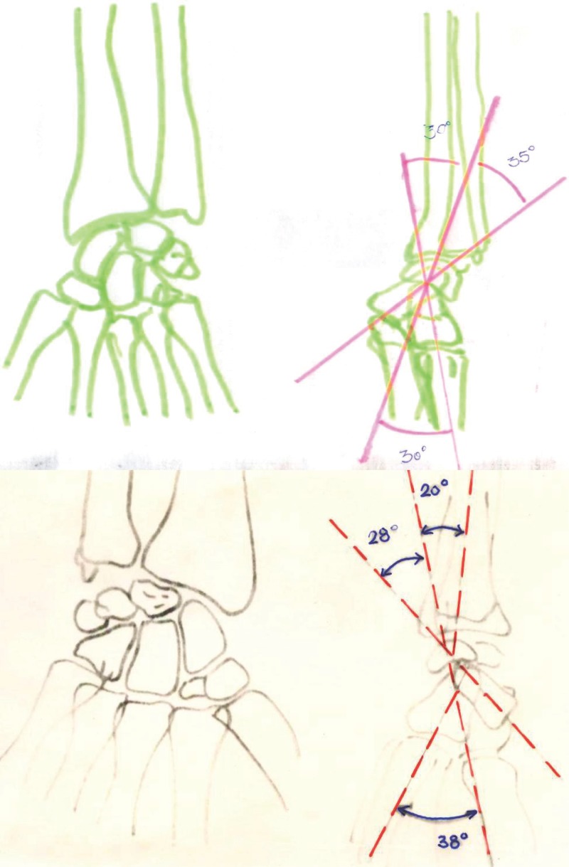
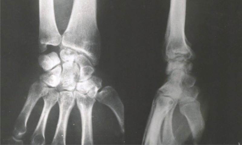
X-ray, CAT and arthroscopy provide “changed anatomical image”–changed the position of the bones and their anatomical relationship (Figure 4). Changed are the articular surfaces and articular spaces (width). Sometimes there is evidence of instability without symptoms. Changed are the position of lunate and scaphoid bone and their properties. Changing is the position of the capitate bone, and thus the axis of the wrist third bone in relation to the axis of the radius bone. Buck–Gramcko (6) talks about “dorsal intercalated segment instability”–DISI. Lunate bone is shifted dorsally, widened joint space between the lunate and scaphoid bone (> 2 mm). Lunate have at the image triangular form, and scaphoid bone is shortened, the lateral image present increased angle between the lunate and scaphoid bones (normal value 46°), increased radio ulnar angle (normal value 10°), changed angle of the lunar and capitatum bone (normal value 0°).
Figure 4.
X–Ray and Schemes “VISI” instability; lunate has been twisted in volar direction; Scaphoid–lunate angle is 28° (less than 30°).
Unlike the dorsal, other type of palmar instability – “Volar Fixed Intercalated segment instability = VISI. Characteristics of the VISI:
Lunate bone twisted in volar direction.
Scaphoid–lunate angle less than 30°
Radio-ulnar angle is negative.
Capitatum–lunate angel is positive.
Lindscheid divides instability in 4 groups.
DISI–Buck Gramcko 1982 (6).
VISI–Buck Gramcko 1982 (6).
Radial wrist translocation.
Ulnar wrist translocation (after destruction) or extirpation of lunate bone.
So that is why Buck-Gramcko in case of lunate bone extirpation replaces the bone with tendon of muscle palmaris longus tendon twisted in form of a ball. (Figure 5)
Figure 5.
28 years after Matti–Risse scaphoid bone surgery asymptomatic “DISI” instability.
2.7. Therapeutic Interventions
Interventions are carried out in three directions:
Rest–immobilization (distension of the ligaments, capsule injury).
Suture or plastics – reconstruction of ledated ligaments.
In case of DISI and VISI instability and deformation it is necessary to establish shifted central axis caused by the shift of lunate bone. Arthrodesis is recommended – lunate bone–capitatum bone (partial arthrodesis). In pseudo arthrodesis scaphoid bone–and recommended is spongioplastics by Matt–Russo–procedure.
wrist arthrodesis; Radio–Scapho–Lunar arthrodesis; Radio–Scapho–arthrodesis; Radio lunar arthrodesis.
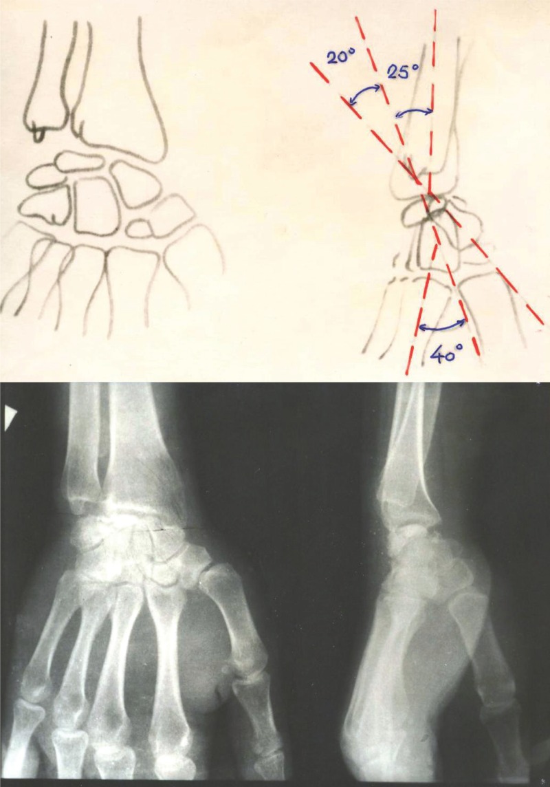
Surgical treatment of acute injury is best performed within 3 weeks of the primary injury. Partial intercarpal fusion is technically demanding and is always associated with loss of range of motion. Fusion of certain carpal bones fails, especially fusion between the scaphoid and the lunate bone, because both bones have a poor blood supply (1). The determining factors in making treatment decisions are: the presence or absence of articular changes, injury chronicity, tissue quality, the possibility of reduction of the deformity (4) (Figure 3).
Figure 3.
X ray and Scheme “VISI” instabilitiy–lunate is rotated in volar direction, scaphoid–lunate angle is 20° (less than 30°).
3. INSTABILITY IN OUR PRACTICE
In our practice in case of old fractures or insufficient healing dominated “VISI” instability (7). From our practice we provided several specific examples.
4. CONCLUSION
Carpal instability may be the result of various injuries of the wrist and is essential in the differential diagnosis of chronic pain in this region. In the past decade was collected a lot of knowledge about these injuries, but still much remains unclear. Diagnosis and treatment are generally difficult and require special skills. Today, the authors prefer the method of clinical examination and X-ray diagnostics, but most patients are then refer to arthroscopy to confirm the diagnosis and exclude other pathologies. Treatment depends on the patient’s symptoms. For those with mild disorder with preserved range of motion by 80% and grip strength surgical treatment is not indicated. For those with serious problems of acute scafo-lunate dissociation (within six weeks of injury), direct access and the dorsal capsule desis are recommended. Finally, in patients with secondary arthritis is performed “rectangle fusion” with scaphoid bone excision or total wrist arthrodesis, depending on the severity of arthritis or the patient’s personal requirements.
CONFLICT OF INTEREST
None declared.
REFERENCES
- 1.Waldram M. Wrist instability. [April 18, 2012];Trauma [serial on the Internet] 1999 Apr;1(2):133–142. Available from: Academic Search Complete. [Google Scholar]
- 2.Fisk GR. Carpal Instability and the Fractured Scaphoid. Ann. Royal Coll Surg Engl. 1970;48:63–76. [PMC free article] [PubMed] [Google Scholar]
- 3.http://www.mef.unizg.hr/ortopedija/predavanja/Nestabilnost%20rucja.pdf . 18.4.12.
- 4.http://www.fimnet.fi/sjs/articles/SJS42008-324.pdf . 18.4.12.
- 5.Gelberman R, Cooney W, Szabo R. Carpal Instability. [April 18, 2012];Journal Of Bone & Joint Surgery, American Volume [serial on the Internet] 2000 Apr;82(4):577. Available from: Academic Search Complete. [Google Scholar]
- 6.Buck-Gramcko D. Instabilitet Des Handgelenes in nigst, H Fracturen, Luxationen Und Dissozitationed Der Karpalknochen, Hipokrates, Stuttgart. 1982:175–183. [Google Scholar]
- 7.Stanley JK, Trail IA. J Bone Joint Surg Br. 1994 Sep;76(5):691–700. [PubMed] [Google Scholar]
- 8.Kauer JMG. Funkcional anatomy of the wrist. Clin. Orthop. 1980;149:9–20. [PubMed] [Google Scholar]
- 9.Kuhlmann JN. Experimentelle untersuchungen zur Stabilität nd instabilität des karpus (Nigst, H.., fracturen, luxationene und dissoziationen der karpal knochen. Stuttgart: Hippokrates; 1982. pp. 185–201. [Google Scholar]
- 10.Martini AK, Cotta H. Handgelenk Instabilität – diagnose und therapie. Georg Thieme verlag Stuttgart – New York. Akt traumat. 1989;19:287–293. [PubMed] [Google Scholar]
- 11.Muminagić S. Kienb”ck desease – Wrist instability mini SICOT. Budimpešta: 1991. [Google Scholar]


