

Abstract
BACKGROUND—Removal of bovine serum from organ culture medium is necessary because of the variability in serum composition and the potential risk of infection. Two specific endothelial cell media (F99 and Endothelial-SFM) were compared with the routinely used medium MEM for their use in serum free cultivation of human corneal endothelial cells (HCEC) and donor corneas. METHODS—HCEC were incubated in three test media with or without increasing serum content and a growth assay was performed. Seven pairs of donor corneas were cultured in each of three media for 3 weeks, one cornea with serum supplementation and one without. Endothelial cell density was determined once each week. Trypan blue staining of the endothelium and vital staining of keratocytes was performed after 3 weeks. RESULTS—All three media promoted proliferation of cultured HCEC when supplemented with serum. Endothelial cell density of donor corneas was comparable after 3 weeks of cultivation in the different media. Only corneas cultured in medium MEM without serum exhibited a higher endothelial cell loss. Trypan blue staining of the endothelium after cultivation revealed the lowest number of damaged cells on corneas cultured in the medium Endothelial-SFM. The highest densities of keratocytes were found in corneas cultured in Endothelial-SFM and the lowest densities occurred after culture in MEM. CONCLUSION—After incubation in Endothelial-SFM even under serum free conditions corneas were found to be of higher quality with respect to endothelial cell survival, cell membrane integrity, and keratocyte density. This medium may replace MEM, which is routinely used in European eye banks but requires supplementation with serum.
Full Text
The Full Text of this article is available as a PDF (135.6 KB).
Figure 1 .
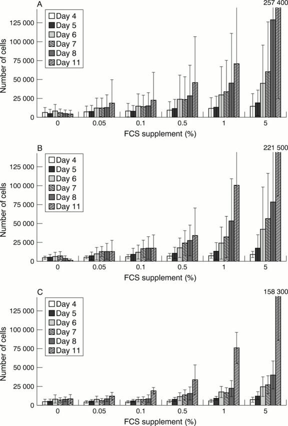
Proliferation of isolated human corneal endothelial cells during culture in medium MEM (A), F99 (B), and Endothelial-SFM (C). Cells were seeded on six well plates (2000 cells/well) and cultured using the indicated medium without serum supplementation or in the presence of 0.05%, 0.1%, 0.5%, 1%, or 5% FCS, respectively. The number of cells was determined after 4, 5, 6, 7, 8, and 11 days of culture. Indicated cell amounts are means (SD) of six independent experiments.
Figure 2 .

Endothelial cell loss of donor corneas during organ culture in medium MEM, F99, and Endothelial-SFM (SFM) with or without supplementation of 2% FCS. Cell densities were determined at the beginning of organ culture and then every week. Indicated are the means (SD) for the decrease in endothelial cell density determined from seven corneas.
Figure 3 .

| p Value | Significance | |
| MEM with FCS versus SFM with FCS | 0.036 | Yes |
| MEM without FCS versus SFM without FCS | 0.003 | Yes |
| MEM with FCS versus F99 with FCS | 0.260 | No |
| MEM without FCS versus F99 without FCS | 0.012 | Yes |
| F99 with FCS versus SFM with FCS | 0.132 | No |
| F99 without FCS versus SFM without FCS | 0.018 | Yes |
| SFM with FCS versus SFM without FCS | 0.943 | No |
| MEM with FCS versus MEM without FCS | 0.100 | No |
| F99 with FCS versus F99 without FCS | 1.000 | No |
Selected References
These references are in PubMed. This may not be the complete list of references from this article.
- Aboalchamat B., Engelmann K., Böhnke M., Eggli P., Bednarz J. Morphological and functional analysis of immortalized human corneal endothelial cells after transplantation. Exp Eye Res. 1999 Nov;69(5):547–553. doi: 10.1006/exer.1999.0736. [DOI] [PubMed] [Google Scholar]
- Barisani-Asenbauer T., Kaminski S., Schuster E., Dietrich A., Biowski R., Lukas J., Gosch-Baumgartner I. Impact of growth factors on morphometric corneal endothelial cell parameters and cell density in culture-preserved human corneas. Cornea. 1997 Sep;16(5):537–540. [PubMed] [Google Scholar]
- Bednarz J., Teifel M., Friedl P., Engelmann K. Immortalization of human corneal endothelial cells using electroporation protocol optimized for human corneal endothelial and human retinal pigment epithelial cells. Acta Ophthalmol Scand. 2000 Apr;78(2):130–136. doi: 10.1034/j.1600-0420.2000.078002130.x. [DOI] [PubMed] [Google Scholar]
- Bourne W. M., Doughman D. J., Lindstrom R. L., Kolb M. J., Mindrup E., Skelnik D. Increased endothelial cell loss after transplantation of corneas preserved by a modified organ-culture technique. Ophthalmology. 1984 Mar;91(3):285–289. doi: 10.1016/s0161-6420(84)34306-8. [DOI] [PubMed] [Google Scholar]
- Bourne W. M. Endothelial cell survival on transplanted human corneas preserved at 4 C in 2.5% chondroitin sulfate for one to 13 days. Am J Ophthalmol. 1986 Sep 15;102(3):382–386. doi: 10.1016/0002-9394(86)90015-2. [DOI] [PubMed] [Google Scholar]
- Duffy P., Wolf J., Collins G., DeVoe A. G., Streeten B., Cowen D. Letter: Possible person-to-person transmission of Creutzfeldt-Jakob disease. N Engl J Med. 1974 Mar 21;290(12):692–693. [PubMed] [Google Scholar]
- Engelmann K., Böhnke M., Friedl P. Isolation and long-term cultivation of human corneal endothelial cells. Invest Ophthalmol Vis Sci. 1988 Nov;29(11):1656–1662. [PubMed] [Google Scholar]
- Engelmann K., Friedl P. Growth of human corneal endothelial cells in a serum-reduced medium. Cornea. 1995 Jan;14(1):62–70. [PubMed] [Google Scholar]
- Engelmann K., Friedl P. Optimization of culture conditions for human corneal endothelial cells. In Vitro Cell Dev Biol. 1989 Nov;25(11):1065–1072. doi: 10.1007/BF02624143. [DOI] [PubMed] [Google Scholar]
- Engelmann K., Sobottka Ventura A., Drexler D., Staude H. J. A sensitive method for testing the quality of organ culture media and of individual medium components in a cornea bank. Graefes Arch Clin Exp Ophthalmol. 1998 Apr;236(4):312–319. doi: 10.1007/s004170050084. [DOI] [PubMed] [Google Scholar]
- Heckmann J. G., Lang C. J., Petruch F., Druschky A., Erb C., Brown P., Neundörfer B. Transmission of Creutzfeldt-Jakob disease via a corneal transplant. J Neurol Neurosurg Psychiatry. 1997 Sep;63(3):388–390. doi: 10.1136/jnnp.63.3.388. [DOI] [PMC free article] [PubMed] [Google Scholar]
- Litzkas P., Jha K. K., Ozer H. L. Efficient transfer of cloned DNA into human diploid cells: protoplast fusion in suspension. Mol Cell Biol. 1984 Nov;4(11):2549–2552. doi: 10.1128/mcb.4.11.2549. [DOI] [PMC free article] [PubMed] [Google Scholar]
- Markus H. S., Duchen L. W., Parkin E. M., Kurtz A. B., Jacobs H. S., Costa D. C., Harrison M. J. Creutzfeldt-Jakob disease in recipients of human growth hormone in the United Kingdom: a clinical and radiographic study. Q J Med. 1992 Jan;82(297):43–51. [PubMed] [Google Scholar]
- Møller-Pedersen T., Møller H. J. Viability of human corneal keratocytes during organ culture. Acta Ophthalmol Scand. 1996 Oct;74(5):449–455. doi: 10.1111/j.1600-0420.1996.tb00597.x. [DOI] [PubMed] [Google Scholar]
- Pels E., Schuchard Y. Organ-culture preservation of human corneas. Doc Ophthalmol. 1983 Dec 15;56(1-2):147–153. doi: 10.1007/BF00154722. [DOI] [PubMed] [Google Scholar]
- Poole C. A., Brookes N. H., Clover G. M. Keratocyte networks visualised in the living cornea using vital dyes. J Cell Sci. 1993 Oct;106(Pt 2):685–691. doi: 10.1242/jcs.106.2.685. [DOI] [PubMed] [Google Scholar]
- Salla S., Redbrake C., Becker J., Reim M. Remarks on the vitality of the human cornea after organ culture. Cornea. 1995 Sep;14(5):502–508. [PubMed] [Google Scholar]
- Schultz G., Cipolla L., Whitehouse A., Eiferman R., Woost P., Jumblatt M. Growth factors and corneal endothelial cells: III. Stimulation of adult human corneal endothelial cell mitosis in vitro by defined mitogenic agents. Cornea. 1992 Jan;11(1):20–27. doi: 10.1097/00003226-199201000-00003. [DOI] [PubMed] [Google Scholar]
- Singh G., Böhnke M., von-Domarus D., Draeger J., Lindstrom R. L., Doughman D. J. Vital staining of corneal endothelium. Cornea. 1985;4(2):80–91. [PubMed] [Google Scholar]