

Abstract
The human life-history is characterized by long development and introduction of new developmental stages, such as childhood and adolescence. The developing brain had important role in these life-history changes because it is expensive tissue which uses up to 80% of resting metabolic rate (RMR) in the newborn and continues to use almost 50% of it during the first 5 postnatal years. Our hominid ancestors managed to lift-up metabolic constraints to increase in brain size by several interrelated ecological, behavioral and social adaptations, such as dietary change, invention of cooking, creation of family-bonded reproductive units, and life-history changes. This opened new vistas for the developing brain, because it became possible to metabolically support transient patterns of brain organization as well as developmental brain plasticity for much longer period and with much greater number of neurons and connectivity combinations in comparison to apes. This included the shaping of cortical connections through the interaction with infant's social environment, which probably enhanced typically human evolution of language, cognition and self-awareness. In this review, we propose that the transient subplate zone and its postnatal remnant (interstitial neurons of the gyral white matter) probably served as the main playground for evolution of these developmental shifts, and describe various features that makes human subplate uniquely positioned to have such a role in comparison with other primates.
Keywords: cerebral cortex, neuron number, life-history, metabolic cost, subplate zone
Introduction
We humans have large brains, and flatter ourselves to be smart. Accordingly, we are prone to think that “bigger is better” and to assume that larger (i.e., more encephalized) brains should have larger computational and cognitive abilities (for a comprehensive and historical review, see Herculano-Houzel, 2009, 2011a,b, 2012a). Some recent versions of this notion assume that improved cognition does not depend on relative brain size (i.e., the level of encephalization), but simply correlates with absolute brain size (Deaner et al., 2007) or with absolute numbers of cortical neurons and their connections and synapses (Roth and Dicke, 2005). However, if advantages of higher encephalization or increased brain size are so obvious, why big brains are so rare (Parker, 1990)? To answer this paradox, one should obtain more detailed knowledge on neural scaling rules in various mammalian orders (Herculano-Houzel, 2011a,b), as well as ask what the costs of encephalization are and how they can be afforded (Foley and Lee, 1991).
Total number of neurons is more important than brain size per se
In a series of recent studies, starting with invention of new quantitative method for comparative analysis of cell and neuron numbers (Herculano-Houzel and Lent, 2005), it was clearly revealed that neural scaling rules evolved differently in different mammalian orders, such as rodents and lagomorphs (Herculano-Houzel et al., 2006, 2011; Herculano-Houzel, 2007), insectivores (Sarko et al., 2009), and primates (Herculano-Houzel et al., 2007; Gabi et al., 2010) including great apes (Herculano-Houzel and Kaas, 2011) and humans (Azevedo et al., 2009; Herculano-Houzel, 2009). These studies pointed out that, in terms of neuronal numbers, the human brain is linearly scaled-up primate brain and that our superior cognitive abilities might simply reflect the largest total number of neurons in the human brain (Herculano-Houzel, 2009, 2012a,b). It seems that basic primate advantage consists in packing more neurons in the same brain volume, thus avoiding prohibitively large increase in brain size (Herculano-Houzel, 2012a,b). In addition, human brains have ~3 times more brain neurons than gorillas and orangutans (Herculano-Houzel and Kaas, 2011). Another important finding concerns the coordinate increase in numbers of neurons in the cerebral cortex and cerebellum and the fact that the vast majority of all brain neurons are found in these two structures (Herculano-Houzel, 2009, 2010, 2011b, 2012a), thus supporting previous findings on cerebral-cerebellar co-evolution (Whiting and Barton, 2003; Ramnani, 2006; Ramnani et al., 2006; Balsters et al., 2010). Therefore, it has been proposed that “the larger the number of neurons in excess of that required to operate the body, the more complex and flexible the behavior of an animal can be expected to be, and thus the larger its cognitive abilities” (Herculano-Houzel, 2012a, p. 336).
The brain is expensive tissue, and only mothers and infants of a “chosen primate” can afford to grow it beyond ordinary expectations
How much of the resting metabolic rate (RMR) is spend to maintain the adult brain? While most mammals expend 3–4% of RMR on brain metabolism (Mink et al., 1981; Armstrong, 1983, 1990), anthropoid primates spend about 8% of RMR to maintain their brains (Armstrong, 1983, 1990; Hofman, 1983a,b; Martin, 1983; Leonard and Robertson, 1992; Genoud, 2002). While the large brain-body-mass ratio of humans (the adult human brain is 2% of the body's mass) is not associated with elevations in RMR (Leonard and Robertson, 1992, 1994), adult humans nevertheless expend two to three times more energy on brain metabolism than other primates, that is 20–25% of RMR (Passmore and Durnin, 1955; Kety, 1957; Holliday, 1986; Aiello and Wheeler, 1995; Leonard et al., 1996; Rolfe and Brown, 1997; Genoud, 2002). These human brain costs are even more impressive during childhood, because the brain consumes roughly 87% of RMR in the newborn, and 44% in a 5 year old child (Holliday, 1986). In comparison to the neonate chimpanzee, the cost of the human neonate brain is significantly greater, and by the age of 5 years these costs are 3 times as great (Foley and Lee, 1991). Such energetic costs seem also to exert a selective pressure toward metabolically efficient neural morphology, that is, metabolically efficient patterning of dendritic arborizations (Wen and Chklovskii, 2008), neural codes (Levy and Baxter, 1996; Balasubramanian et al., 2001), and brain wiring patterns (Chen et al., 2006).
Positive pleiotropic gene effects on relative brain and body growth occur during prenatal and early postnatal periods, because genes affecting both traits generally do so during fetal and early postnatal growth, when both brain and body size are growing rapidly (Riska and Atchley, 1985).The fetal brain at any stage of development constitutes a markedly larger proportion of total fetal weight in primates than in other mammals (Sacher, 1982), and this difference is still observable in neonates (Martin, 1983). However, this difference is no longer clear in comparisons among adults, due to differential postnatal changes in different mammals (Martin, 1983). This points to the crucial importance of brain development (Martin, 1996). For example, evolutionary shifts in brain development lead to differences in development of social behavior and cognition even between such closely related species such as chimpanzees and bonobos (Wobber et al., 2010).
The growth of the brain significantly depends upon energetic and hence ecological conditions (Martin, 1983; Foley and Lee, 1991); as succintly stated by Foley and Lee (1991, p. 223): “whatever selective pressures there may be driving the size of the brain up, these are satisfied only in the context of there being sufficient energy.” Having a large brain imposes additional energetic costs on both the infant and the mother; the mother can derive that energy either from the incorporation of higher quality food, from feeding for longer each day, or from maintaining lactation over a longer period (Foley and Lee, 1991). Thus, the evolution of a large brain requires that energetic constraints are lifted (Armstrong, 1983; Hofman, 1983a,b, 1993; Martin, 1983, 1996; Foley and Lee, 1991; Leonard and Robertson, 1992, 1994; Aiello and Wheeler, 1995; Leonard et al., 2003; Isler and van Schaik, 2006a,b, 2009).
Some recent evidence suggests that the metabolic cost may be an even more limiting factor to brain expansion than previously suspected (Herculano-Houzel, 2012b). Namely, the estimated glucose use per neuron is remarkably constant, varying only by 40% across the six species of rodents and primates, including humans (Herculano-Houzel, 2011c). Thus, it seems that the brain energy budget per neuron is fixed across species and brain sizes and that the total metabolic cost of a brain is a simple, direct function of its number of neurons (Herculano-Houzel, 2011c). These findings clearly suggest that neuronal metabolism imposes a series of constraints upon brain structure, function, and evolution (Herculano-Houzel, 2011c, 2012b; Fonseca-Azevedo and Herculano-Houzel, 2012). The metabolic constraints upon brain scaling in evolution are imposed by absolute number of neurons, because adding neurons to the brain comes at a sizable cost of 6 kcal/d per billion neurons (Herculano-Houzel, 2011c).
Three major hypotheses have been proposed to explain how larger brains are afforded among mammalian species (see Jones and MacLarnon, 2004, for a comprehensive review): direct metabolic constraint hypothesis (Armstrong, 1983; Hofman, 1983a,b); the expensive tissue hypothesis (Aiello and Wheeler, 1995); and the maternal energy hypothesis (Martin, 1981, 1983, 1996, 2007; Martin et al., 2005). None of these hypotheses has the general applicability in multiple mammalian clades with different evolutionary histories (Jones and MacLarnon, 2004), and there are several strategies for meeting the energetic demands of encephalization which can be manifested differentially across taxa (Barrickman and Lin, 2010). However, at least in the case of large-brained apes and humans, the maternal energy hypothesis seems to be well supported by the available evidence (Martin, 1996; Isler and van Schaik, 2006a,b, 2009; Isler et al., 2008). This hypothesis proposes that the brain size is constrained by the amount of energy that a mother can provide during the early stages of her offspring's ontogeny (Martin, 1996, 2007). It should be noted that such a primary link between the mother's metabolic capacity and the developing brain of her offspring allows other variables to influence ultimate adult brain size (Martin, 1996). In addition, there may be no very tight relationship between relative brain size and specific behavioral capacities, and an increase in brain size may be advantageous in a diffuse fashion, i.e., may have some kind of permissive or promotive influence with respect to the evolution of cognition (Martin, 1996).
The encephalization is also associated with prolonged duration of most life-history stages, especially in primates (Sacher and Staffeldt, 1974; Harvey and Clutton-Brock, 1985; Barton, 1999; Kappeler and Pereira, 2003; Leigh, 2004; Barrickman et al., 2008) including humans (Bogin, 1997, 1999, 2009; Leigh, 2001). At least three changes in developmental timing occurred during the evolution of human encephalization: extended brain growth, retarded postnatal body growth, and a derived brain growth allometry (Vinicius, 2005). The human brain achieves its final size more by lengthening the time of growth than by adopting an unusual rate of growth (Passingham, 1985). The prolonged period of growth in humans may be partly an adaptation to limit the already high total and brain energy requirements during childhood (Leonard and Robertson, 1992). Others have suggested that selection has acted to decrease human somatic growth rates during childhood and juvenility (in comparison to chimpanzees), to help fuel the energy-expensive brain and to allow more time for increased cognitive development with lower body-maintenance costs (Walker et al., 2006).
So, how our evolving ancestors have solved the above mentioned energetic challenges? Obviously, there were a number of step-wise changes, stretching perhaps over last 2 million years, i.e., during the evolution of the genus Homo. One part of the solution seems to be a significant change in dietary and foraging habits, as humans have a much higher quality diet than expected for their size or their resting metabolic needs (Leonard and Robertson, 1994, 1997; Fish and Lockwood, 2003). Turning to animal source foods, such as meat, as a routine dietary component probably represented an important step (Milton, 1999, 2003). However, it seems that a diet relying solely on consumption of raw food was not sufficient to remove this metabolic constraint on the increase of brain size—as documented in a recent study, the largest great apes cannot afford both a large body and a larger number of brain neurons (Fonseca-Azevedo and Herculano-Houzel, 2012). The use of fire and the invention of cooking might have a substantial role, because the cooking increases enormously the energy yield of foods and the speed with which they are consumed (Carmody and Wrangham, 2009; Carmody et al., 2011). While raw meat increased the caloric content of the diet of early hominids (Milton, 1999), the cooked meat is easier to chew and has a higher caloric yield (Carmody et al., 2011). In fact, as the metabolic cost is limiting enough to impose tradeoffs in brain evolution (Fonseca-Azevedo and Herculano-Houzel, 2012), the invention of cooking food was probably necessary to overcome such a metabolic limitation in the human lineage (Wrangham et al., 1999; Wobber et al., 2008; Carmody and Wrangham, 2009; Carmody et al., 2011). It should be also noted that there is evidence of up-regulation of genes related to energy metabolism in human evolution (Grossman et al., 2001; Cáceres et al., 2003; Uddin et al., 2004).
Another part of the solution seems to be represented by profound changes in the human life-history (Bogin, 1997, 1999, 2001, 2009; Hawkes et al., 1998; Kaplan et al., 2000; Crews, 2003; Leigh, 2004; Gurven and Walker, 2006; Walker et al., 2006). There are several hypotheses on the evolution of human life-history, such as the grandmother hypothesis (Hawkes et al., 1998), the embodied capital hypothesis (Kaplan et al., 2000), the reserve capacity hypothesis (Crews, 2003; Larke and Crews, 2006), and the reproductive fitness hypothesis (Bogin, 1997, 1999, 2009). Briefly, primates and other social mammals have three postnatal life history stages: infancy, juvenile and adult (Pereira and Fairbanks, 1993). However, human life history is characterized by the addition of childhood, adolescence, and grandmotherhood (postmenopausal stage) as biologically, behaviorally, and mathematically definable stages of the life cycle (Bogin, 1997, 1999; Hawkes et al., 1998). The transition from infancy (birth to 30–36 months) to childhood is characterized by weaning and the completion of deciduous tooth eruption (Bogin, 1999, 2001). During the childhood, older members of the social group acquire, prepare, and provision foods to children, and this style of cooperative care represents a major evolutionary invention in the human life-history (Bogin, 1999, 2009). The adolescence includes the years of postpubertal growth (10–18 years for girls, 12–21 years for boys) (Bogin, 1999, 2001).
It is important to note that the childhood and adolescence stages of human life history evolved due to the selective advantages for increased fertility and reproductive fitness of mothers (Bogin, 1999, 2001, 2009), while the benefits of these stages for increased brain growth and learning are important, but secondary, outcomes (Bogin, 2009). In summary, the human species has more life stages than any other mammal and more time for growth and development than any primate (Bogin, 2009). Another important evolutionary novelty in human life-history is that human food provisioning and care to children and their mothers goes beyond the cooperative breeding of other mammals—humans use biological relationships and also marriage, systems of economic exchange, political power structure, and gender-role construction (Bogin, 2009). In other words, human life-history is culturally patterned (Bogin, 2001, 2009; Crews, 2003). Such investments of energy and care from prenatal to early adult life stages build a greater level of reserve capacity than found in any other primate (Crews, 2003; Larke and Crews, 2006; Bogin, 2009).
The subplate is critically involved in the ontogenesis of the human cerebral cortex
The data reviewed above clearly suggest that the developing brain played significant role in the evolution of the human life-history. As the telencephalon and the cerebral cortex represent by far the largest part of the human brain, we here focus on the potential evolutionary role of the transient subplate zone, because it is critically involved in the development of the primate and human cerebral cortex (Bystron et al., 2008) and it reached a peak of its evolutionary prominence in the human brain (Kostovic and Rakic, 1990; Molnár et al., 2006; Rakic, 2006; Bystron et al., 2008). The role of the subplate in the development and plasticity of the cerebral cortex has been already well described in a number of excellent reviews (Allendoerfer and Shatz, 1994; Kostović and Judaš, 2002, 2006, 2007, 2010; Kanold and Shatz, 2006; Molnár et al., 2006; Rakic, 2006, 2009; Bystron et al., 2008; Kanold and Luhmann, 2010; Clowry et al., 2010; Judaš, 2011). Therefore, we will here only briefly review those aspects of the human subplate which are directly relevant for understanding of our present thesis. As the subplate development in the human brain has also been extensively illustrated in our previous publications (Kostovic and Rakic, 1980, 1990; Kostović and Judaš, 2002, 2006, 2007, 2010; Judaš, 2011), we here provide only a few figures aimed to enhance the understanding of our main argument.
The subplate zone was first described in the human fetal brain (Kostović and Molliver, 1974; Judaš et al., 2010a; see, for a comprehensive historical review). The subplate is the largest transient compartment of the fetal neocortical anlage (see Judaš, 2011, for a comprehensive review). The human subplate develops between 13 and 15 postconceptional weeks (PCW), remains the largest compartment of the neocortical anlage between 15 and 30 PCW, and begins slowly to disappear toward the end of gestation and during the early postnatal period (Figure 1). The developmental peak of the subplate is reached during midgestation, when it is about four times thicker than the cortical plate (Figure 2). It should be noted that the subplate is still present in the newborn brain during the period when various corticocortical connections continue to develop (Figure 3). Finally, many subplate neurons survive postnatally and eventually transform into interstitial neurons of the subcortical (gyral) white matter of the adolescent and adult brain (Figures 4, 5) (Kostovic and Rakic, 1980, 1990; Judaš et al., 2010b). While the dissolution of subplate begins during the last third of gestation, it remains present (as recognizable architectonic compartment) under the prefrontal and other association cortices up to 6 postnatal months (Kostovic and Rakic, 1990). It should be noted with a great regret that there are no data available on the subplate of great apes; in fact, there are no histological data on any aspect of prenatal cortical development in great apes.
Figure 1.
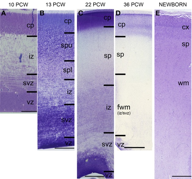
Laminar development of human telencephalon from 10 postconceptional weeks (PCW) to newborn. The layers are transient and their appearance changes with changes in neurogenetic events. The subplate starts to develop around 13 PCW, reaches the peak of its development between 22 and 24 PCW, and starts to resolve around 34 PCW. In the newborn brain, the subplate remains during the first year, when the subplate disappears as a zone but its neurons become incorporated into the subcortical white matter as so-called interstitial neurons. cp, cortical plate; sp, subplate zone; iz, intermediate zone; svz, subventricular zone; vz, ventricular zone. Bar = 100 μm (A), 250 μm (B), 1 mm (C–E).
Figure 2.
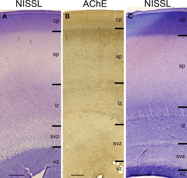
Lamination of frontal (A), mid-central (B) and occipital (C) region of human telencephalon at the peak of subplate development (22–24 PCW), as revealed by Nissl staining and acetylcholinesterase (AChE) histochemistry. At the peak of subplate development (22–24 PCW), subplate zone is the largest compartment of the human telencephalon. It is the place of intense synaptic activity and “waiting” compartment for the thalamocortical fibers (dark band below cp). Note that there are regional differences in the lamination between frontal and occipital region. cp, cortical plate; sp, subplate zone; iz, intermediate zone; svz, subventricular zone; vz, ventricular zone. Bar = 1 mm.
Figure 3.
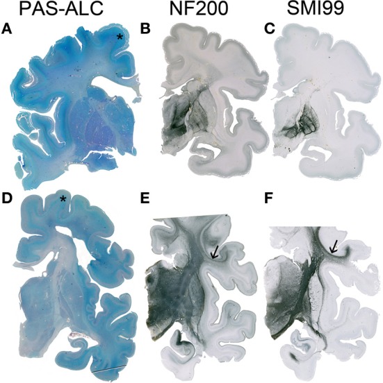
Although the resolution of the subplate zone starts after 34 PCW, the subplate remains visible as a major component of the telecephalic wall (asterisk in A,D). In the human telencephalon, cortico-cortical connections are still not developed (B) or myelinated (C) at 33 PCW, while at 40 PCW substantial development and myelination of cortico-cortical fiber can be observed (arrow in E,F). A–C 34 PCW, D–F 40 PCW.
Figure 4.
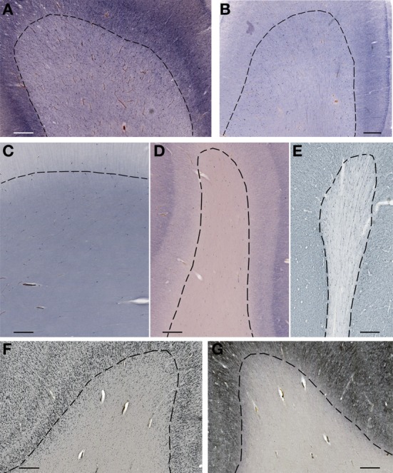
Subplate and white matter interstitial neurons stained for NOS (NADPH-diaphorase-stained neurons in A–D), MAP2 (E,G) and NeuN (F) are visible throughout the subplate (A,B) and the white matter (C–G). Note that subplate/white matter interstitial neurons are numerous even after the first year of life, when subplate zone disappears. (A), 37 PCW; (B), 13 days; (C), 12 years; (D), 57 years; (E,G), 13 months; (F), 51 years. Bar = 1 mm.
Figure 5.
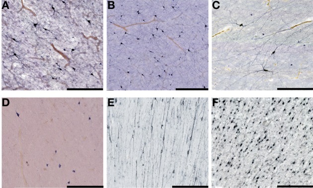
Higher magnification view of subplate and white matter interstitial neurons displayed in panels A–F of the Figure 4 and stained for NOS (NADPH-diaphorase-stained neurons in A–D), MAP2 (E) and NeuN (F). Note that dendritic arborizations of subplate/interstitial neurons continue to grow and develop even after the disapearance of the subplate zone during the first year of life (compare (A and B with C). (A), 37 PCW; (B), 13 days; (C), 12 years; (D), 57 years; (E), 13 months; (F), 51 years. Bar = 0.5 mm.
The subplate contains numerous neurons of various morphological types (Mrzljak et al., 1988, 1990, 1992) and molecular phenotypes, including differentiated projection (glutamatergic) neurons and local (GABA and peptidergic) interneurons (Judaš et al., 1999, 2010b; Judaš, 2011). It also serves as a waiting compartment for growing cortical afferents (Rakic, 1977; Kostovic and Rakic, 1990). Various afferent fibers sequentially grow into the subplate, establish temporary synaptic circuits, and “wait” in the subplate for several months before relocating into their final target, the cortical plate (Kostović and Goldman-Rakic, 1983; Krmpotić-Nemanić et al., 1983; Kostovic and Rakic, 1984, 1990; Kostović, 1986). After 28 PCW, waiting associative and commissural pathways are major constituents of the subplate (Kostovic and Rakic, 1990; Kostović et al., 2008; Kostović and Judaš, 2009). While long corticocortical pathways begin to develop in the early fetal period (Vasung et al., 2010), the development of short corticocortical connections is very protracted and lasts for at least 1 year after birth (Kostović et al., 2012). It should be noted that cortical pyramidal neurons also require about 3 years of postnatal development in order to attain their adult-like size of dendritic arborization (Petanjek et al., 2008).
The subplate is also the major site of synaptogenesis in the midfetal brain (Molliver et al., 1973; Kostovic and Rakic, 1990) and contains diverse and transient neuronal circuits which represent a neurobiological basis for transient electrophysiological and behavioral phenomena in fetuses and early preterm infants (Kostović and Judaš, 2002, 2006, 2007, 2010). Although the onset of cortical synaptogenesis is an early fetal event (Molliver et al., 1973; Kostovic and Rakic, 1990), it should be noted that cortical synaptogenesis is predominantly postnatal process and that synaptic overproduction and developmental plasticity in the human cortex continue for at least 20 years (Petanjek et al., 2011).
The transformation of the fetal white matter occurs gradually and in parallel with gradual dissolution of the subplate, and continues postnatally (Judaš, 2011). The period spanning the last prenatal month and at least the first postnatal year is characterized by significant fiber-architectonic reorganization at the cortical/white matter interface (Kostović et al., 2012). This reorganization is related to the postnatal persistence of the subplate remnant, the onset of myelination, the appearance of tertiary gyri and sulci, development of short corticocortical connections (Kostović et al., 2012), and probably other factors, such as changes in microvascular network, changes in molecular profile of the extracellular matrix, development of white matter astrocytes, and so forth (Judaš, 2011).
Thus, histogenetic processes in the human fetal and perinatal brain are protracted and significantly overlap (Judaš, 2011), but the subplate represents a playground for the majority of important events during that developmental window. The functional significance of transient fetal circuitry and the pivotal role of the subplate have already been extensively reviewed in both experimental model animals (Allendoerfer and Shatz, 1994; Kanold and Luhmann, 2010) and in humans (Kostović and Judaš, 2006, 2007, 2010; Judaš, 2011). Therefore, it will suffice to point out that the human perinatal and early postnatal period is characterized by simultaneous existence of two separate (but interconnected) types of cortical circuitry organization: (a) transient fetal circuitry, centered at the subplate zone, and (b) immature but progressively developing permanent cortical circuitry, centered at the cortical plate (that is, developing cortical layers I-VI). Thus, the developing human cortex passes through three major early stages of functional development (Kostović and Judaš, 2006, 2007, 2010): (1) initial fetal circuitry which is endogeneously (spontaneously) driven, (2) perinatal dual circuitry (co-existence of endogeneously driven subplate-centered transient circuitry with developing cortical plate-centered permanent circuitry) and (3) postnatally established permanent (externally driven) cortical circuitry (Judaš, 2011).
The subplate as the playground for evolution of cortical development
While the focus of this review is on putative (and relatively recent) evolutionary changes of the subplate in the primate and hominid lineage, it is important to note that the subplate may have a much older phylogenetic origin. As pointed out in several recent studies (Montiel et al., 2011; Wang et al., 2011), there are currently three hypotheses about the phylogenetic origin of subplate neurons: (1) subplate neurons were all already present in the common ancestor of mammals and sauropsids (e.g., Marin-Padilla, 1978; Aboitiz et al., 2005); (2) subplate may be unique to mammals and represent an embryonic adaptation to support development of increasingly complex neocortex (Kostovic and Rakic, 1990; Supér and Uylings, 2001; Molnár et al., 2006); and (3) the subplate in mammals may represent a combination of new and ancestral cell populations (Aboitiz, 1999; Aboitiz et al., 2005; Wang et al., 2011; Montiel et al., 2011). The third hypothesis suggests that, although embryonic subplate cells were present in the common ancestor of both mammals and sauropsids, additional populations of subplate cells evolved in mammals as the neocortex became progressively larger and more complex (Montiel et al., 2011; Wang et al., 2011). As the evolution of the mammalian cortex required the modification of developmental programs, it seems probable that some of these started to rely on novel populations of subplate neurons possibly characterized by different targets of connectivity (Kostovic and Rakic, 1990; Montiel et al., 2011). Thus, it is important to determine if and how the subplate has been altered in distinct mammalian lineages and to perform comparative gene expression profiling studies of subplate neurons in different species (Osheroff and Hatten, 2009; Wang et al., 2010, 2011; Oeschger et al., 2012; Hoerder-Suabedissen et al., 2013). For example, species-specific differences in subplate markers have been described even between rat and mouse (Wang et al., 2011). In addition, in primates, in contrast to rodents, neurons are continuously added to the subplate throughout cortical neurogenesis (Smart et al., 2002; Lukaszewicz et al., 2005; Molnár et al., 2006). Finally, in addition to the increased number of neurons in the human subplate (Kostovic and Rakic, 1990; Smart et al., 2002; Bystron et al., 2008), there is both an increased complexity of subplate cell types (Kostovic and Rakic, 1990; Mrzljak et al., 1988, 1990, 1992; Wang et al., 2010) and subplate arrangements including the superficial vs. deep compartmentalization of human subplate neurons (Wang et al., 2010).
Thus, the available evidence suggests that human subplate contains an increased number of (ancestral and derived) subplate neurons as well as increased diversity of a derived population of subplate neurons. As these neurons are active and therefore metabolically expensive, the potential increase in number of subplate neurons was probably subject to a significant selective pressure due to above described metabolic constraints.
The lift-up of metabolic constraints by hominid ancestors opened new vistas for the developing brain, because it became possible to metabolically support transient patterns of brain organization as well as developmental brain plasticity for much longer period and with much greater number of neurons and connectivity combinations in comparison to apes. We propose that the transient subplate zone and its postnatal remnant (interstitial neurons of the gyral white matter) probably served as the main playground for evolution of these developmental shifts, for the following reasons.
First, as described above, the human brain contains about three times more neurons than the brain of apes. As monkey and human cortical neurons are all generated before birth (Rakic, 2006, 2009; Bystron et al., 2008), and newborn human brain is also significantly larger than that of newborn apes (ca. 350 vs. ca. 200 g), it is logical to conclude that brains of human newborns also contain greatly increased number of neurons in comparison to newborn apes. By extension, even if we assume that apes have proportionately equally developed subplate, humans would still have more numerous subplate neurons. Moreover, that huge number of subplate neurons is actively involved in shaping of cortical circuitry for at least 12 months (Judaš, 2011; Kostović et al., 2012), and large number of subplate neurons survives into adolescence and adulthood as subcortical interstitial neurons (Judaš et al., 2010b). Thus, significantly enlarged number of key players in developmental cortical plasticity is present and metabolically supported to play this game for much longer than in any other primate species.
Second, as also described above, the subplate serves as a “waiting” compartment for numerous contingents of ingrowing cortical afferents. The human subplate contains the largest amount of both subcortical and corticocortical waiting afferents, during the longest developmental period. The subplate is the major site of synaptogenesis and early circuit formation during the prenatal period. Its circuitry also coexists with initial adult-like circuitry during the perinatal period, and its neurons continue to be involved in the development of short corticocortical connections during the first postnatal year (Kostović et al., 2012). Thus, humans become able to sustain an extremely long period of cortical circuitry development, characterized by large overproduction of axonal and dendritic branches, synapses and reorganizational events in response to environmental influences. This includes the shaping of cortical connections through the interaction with infant's social environment, which probably enhanced typically human evolution of language, cognition and self-awareness.
In summary, we propose that life-history changes that enabled the metabolic sustainability of prolonged retention of the subplate also provided the playground for prolonged and more diverse perinatal and early postnatal plastic interactions between the increased number of subcortical and corticocortical afferents and increased number of cortical neurons (including the perinatal co-existence of fetal and adult-like cortical circuitry). This enabled the evolution of new types of modular, areal and connectional organization of the human cerebral cortex, subserving cognition and language. Our proposal is also in agreement with the reserve capacity hypothesis (Crews, 2003; Larke and Crews, 2006) and the reproductive fitness hypothesis (Bogin, 1997, 1999, 2001, 2009), because the increased reserve capacity of human species (in comparison to apes) clearly enables the longer development of the human brain, with significant consequences for learning and socialization as well as plasticity and recovery after brain lesions.
Conflict of interest statement
The authors declare that the research was conducted in the absence of any commercial or financial relationships that could be construed as a potential conflict of interest.
Acknowledgments
This study has been supported by the Croatian Science Foundation (HZZ) Grant no. 09.01/414 to Miloš Judaš. Authors gratefully acknowledge the technical assistance of Danica Budinšćak and Zdenka Cmuk in the preparation of histological slides.
References
- Aboitiz F. (1999). Comparative development of the mammalian isocortex and the reptilian dorsal ventricular ridge. Evolutionary considerations. Cereb. Cortex 9, 783–791 10.1093/cercor/9.8.783 [DOI] [PubMed] [Google Scholar]
- Aboitiz F., Montiel J., Garcia R. R. (2005). Ancestry of the mammalian preplate and its derivatives: evolitonary relicts or embryonic adaptations. Rev. Neurosci. 16, 359–376 [DOI] [PubMed] [Google Scholar]
- Aiello L. C., Wheeler P. (1995). The expensive tissue hypothesis: the brain and the digestive system in human and primate evolution. Curr. Anthropol. 36, 199–221 10.1086/204350 [DOI] [Google Scholar]
- Allendoerfer K. L., Shatz C. J. (1994). The subplate, a transient neocortical structure: its role in the development of connections between thalamus and cortex. Annu. Rev. Neurosci. 17, 185–218 10.1146/annurev.ne.17.030194.001153 [DOI] [PubMed] [Google Scholar]
- Armstrong E. (1983). Relative brain size and metabolism in mammals. Science 220, 1302–1304 10.1126/science.6407108 [DOI] [PubMed] [Google Scholar]
- Armstrong E. (1990). Brains, bodies and metabolism. Brain Behav. Evol. 36, 166–176 10.1159/000115305 [DOI] [PubMed] [Google Scholar]
- Azevedo F. A. C., Carvalho L. R. B., Grinberg L. T., Farfel J. M., Ferretti R. E. L., Leite R. E. P., et al. (2009). Equal numbers of neuronal and nonneuronal cells make the human brain an isometrically scaled-up primate brain. J. Comp. Neurol. 513, 532–541 10.1002/cne.21974 [DOI] [PubMed] [Google Scholar]
- Balasubramanian V., Kimber D., Berry M. J., 3rd. (2001). Metabolically efficient information processing. Neural Comp. 13, 799–815 10.1162/089976601300014358 [DOI] [PubMed] [Google Scholar]
- Balsters J. H., Cussans E., Diedrichsen J., Phillips K. A., Preuss T. M., Rilling J. K., et al. (2010). Evolution of the cerebellar cortex: the selective expansion of prefrontal-projecting cerebellar lobules. Neuroimage 49, 2045–2052 10.1016/j.neuroimage.2009.10.045 [DOI] [PMC free article] [PubMed] [Google Scholar]
- Barrickman N. L., Bastian M. L., Isler K., van Schaik C. P. (2008). Life history costs and benefits of increased brain size: a comparative test using primates. J. Hum. Evol. 54, 568–590 10.1016/j.jhevol.2007.08.012 [DOI] [PubMed] [Google Scholar]
- Barrickman N. L., Lin M. J. (2010). Encephalization, expensive tissues, and energetics: an examination of the relative costs of brain size in Strepsirrhines. Am. J. Phys. Anthropol. 143, 579–590 10.1002/ajpa.21354 [DOI] [PubMed] [Google Scholar]
- Barton R. A. (1999). The evolutionary ecology of the primate brain, in Comparative Primate Socioecology, ed Lee P. (Cambridge: Cambridge University Press; ), 167–194 [Google Scholar]
- Bogin B. (1997). Evolutionary hypo-theses for human childhood. Yearb. Phys. Anthropol. 40, 63–89 [Google Scholar]
- Bogin B. (1999). Evolutionary perspective on human growth. Annu. Rev. Anthropol. 28, 109–153 10.1146/annurev.anthro.28.1.109 [DOI] [PubMed] [Google Scholar]
- Bogin B. (2001). The Growth of Humanity. New York, NY: Wiley-Liss [Google Scholar]
- Bogin B. (2009). Childhood, adolescence, and longevity: a multilevel model of the evolution of reserve capacity in human life history. Am. J. Hum. Biol. 21, 567–577 10.1002/ajhb.20895 [DOI] [PubMed] [Google Scholar]
- Bystron I., Blakemore C., Rakic P. (2008). Development of human cerebral cortex: Boulder Committee revisited. Nat. Rev. Neurosci. 9, 110–122 10.1038/nrn2252 [DOI] [PubMed] [Google Scholar]
- Cáceres M., Lachuer J., Zapala M. A., Redmond J. C., Kudo L., Geschwind D. H., et al. (2003). Elevated gene expression levels distinguish human from non-human primate brains. Proc. Natl. Acad. Sci. U.S.A. 100, 13030–13035 10.1073/pnas.2135499100 [DOI] [PMC free article] [PubMed] [Google Scholar]
- Carmody R. N., Weintraub G. S., Wrangham R. W. (2011). Energetic consequences of thermal and nonthermal food processing. Proc. Natl. Acad. Sci. U.S.A. 108, 19199–19203 10.1073/pnas.1112128108 [DOI] [PMC free article] [PubMed] [Google Scholar]
- Carmody R. N., Wrangham R. W. (2009). The energetic significance of cooking. J. Hum. Evol. 57, 379–391 10.1016/j.jhevol.2009.02.011 [DOI] [PubMed] [Google Scholar]
- Chen B. L., Hall D. H., Chklovskii D. B. (2006). Wiring optimization can relate neuronal structure and function. Proc. Natl. Acad. Sci. U.S.A. 103, 4723–4728 10.1073/pnas.0506806103 [DOI] [PMC free article] [PubMed] [Google Scholar]
- Clowry G., Molnár Z., Rakic P. (2010). Renewed focus on the developing human neocortex. J. Anat. 217, 276–288 10.1111/j.1469-7580.2010.01281.x [DOI] [PMC free article] [PubMed] [Google Scholar]
- Crews D. (2003). Human Senescence: Evolutionary and Biocultural Perspectives. Cambridge: Cambridge University Press; 10.1017/CBO9780511542350 [DOI] [Google Scholar]
- Deaner R. O., Isler K., Burkart J., van Schaik C. (2007). Overall brain size, and not encephalization quotient, best predicts cognitive ability across non-human primates. Brain Behav. Evol. 70, 115–124 10.1159/000102973 [DOI] [PubMed] [Google Scholar]
- Fish J. L., Lockwood C. A. (2003). Dietary constraints on encephalization in primates. Am. J. Phys. Anthropol. 120, 171–181 10.1002/ajpa.10136 [DOI] [PubMed] [Google Scholar]
- Foley R. A., Lee P. C. (1991). Ecology and energetics of encephalization in hominid evolution. Phil. Trans. R. Soc. Lond. B 334, 223–232 10.1098/rstb.1991.0111 [DOI] [PubMed] [Google Scholar]
- Fonseca-Azevedo K., Herculano-Houzel S. (2012). Metabolic constraint imposes tradeoff between body size and number of brain neurons in human evolution. Proc. Natl. Acad. Sci. U.S.A. 109, 18571–18576 10.1073/pnas.1206390109 [DOI] [PMC free article] [PubMed] [Google Scholar]
- Gabi M., Collins C. E., Wong P., Torres L. B., Kaas J. H., Herculano-Houzel S. (2010). Cellular scaling rules for the brains of an extended number of primate species. Brain Behav. Evol. 76, 32–44 10.1159/000319872 [DOI] [PMC free article] [PubMed] [Google Scholar]
- Genoud M. (2002). Comparative studies of basal rate of metabolism in primates. Evol. Anthropol. 11, 108–111 10.1002/evan.10070 [DOI] [Google Scholar]
- Grossman L. I., Schmidt T. R., Wildman D. E., Goodman M. (2001). Molecular evolution of aerobic energy metabolism in primates. Mol. Phylogenet. Evol. 18, 26–36 10.1006/mpev.2000.0890 [DOI] [PubMed] [Google Scholar]
- Gurven M., Walker R. (2006). Energetic demand of multiple dependents and the evolution of slow human growth. Proc. R. Soc. B 273, 835–841 10.1098/rspb.2005.3380 [DOI] [PMC free article] [PubMed] [Google Scholar]
- Harvey P. H., Clutton-Brock T. H. (1985). Life history variation in primates. Evolution 39, 559–581 10.2307/2408653 [DOI] [PubMed] [Google Scholar]
- Hawkes K., O'Connell J. F., Blurton Jones N. G., Alvarez H., Charnov E. L. (1998). Grandmothering, menopause, and the evolution of human life histories. Proc. Natl. Acad. Sci. U.S.A. 95, 1336–1339 [DOI] [PMC free article] [PubMed] [Google Scholar]
- Herculano-Houzel S. (2007). Encephalization, neuronal excess, and neuronal index in rodents. Anat. Rec (Hoboken) 290, 1280–1287 10.1002/ar.20598 [DOI] [PubMed] [Google Scholar]
- Herculano-Houzel S. (2009). The human brain in numbers: a linearly scaled-up primate brain. Front. Hum. Neurosci. 3:31 10.3389/neuro.09.031.2009 [DOI] [PMC free article] [PubMed] [Google Scholar]
- Herculano-Houzel S. (2010). Coordinated scaling of cortical and cerebellar numbers of neurons. Front. Neuroanat. 4:12 10.3389/fnana.2010.00012 [DOI] [PMC free article] [PubMed] [Google Scholar]
- Herculano-Houzel S. (2011a). Not all brains are made the same: new views on brain scaling in evolution. Brain Behav. Evol. 78, 22–36 [DOI] [PubMed] [Google Scholar]
- Herculano-Houzel S. (2011b). Brains matter, bodies maybe not: the case for examining neuron numbers irrespective of body size. Ann. N.Y. Acad. Sci. 1225, 191–199 [DOI] [PubMed] [Google Scholar]
- Herculano-Houzel S. (2011c). Scaling of brain metabolism with a fixed energy budget per neuron: implications for neuronal activity, plasticity and evolution. PLoS ONE 6:e17514 10.1371/journal.pone.0017514 [DOI] [PMC free article] [PubMed] [Google Scholar]
- Herculano-Houzel S. (2012a). Neuronal scaling rules for primate brains: the primate advantage. Prog. Brain Res. 195, 325–340 [DOI] [PubMed] [Google Scholar]
- Herculano-Houzel S. (2012b). The remarkable, yet not extraordinary, human brain as a scaled-up primate brain and its associated cost. Proc. Natl. Acad. Sci. U.S.A. 109(Suppl. 1), 10661–10668 [DOI] [PMC free article] [PubMed] [Google Scholar]
- Herculano-Houzel S., Collins C. E., Wong P., Kaas J. H. (2007). Cellular scaling rules for primate brains. Proc. Natl. Acad. Sci. U.S.A. 104, 3562–3567 10.1073/pnas.0611396104 [DOI] [PMC free article] [PubMed] [Google Scholar]
- Herculano-Houzel S., Kaas J. H. (2011). Gorilla and orangutan brains conform to the primate cellular scaling rules: implications for human evolution. Brain Behav. Evol. 77, 33–44 10.1159/000322729 [DOI] [PMC free article] [PubMed] [Google Scholar]
- Herculano-Houzel S., Lent R. (2005). Isotropic fractionator: a simple, rapid method for the quantification of total cell and neuron numbers in the brain. J. Neurosci. 25, 2518–2521 10.1523/JNEUROSCI.4526-04.2005 [DOI] [PMC free article] [PubMed] [Google Scholar]
- Herculano-Houzel S., Mota B., Lent R. (2006). Cellular scaling rules for rodent brains. Proc. Natl. Acad. Sci. U.S.A. 103, 12138–12143 10.1073/pnas.0604911103 [DOI] [PMC free article] [PubMed] [Google Scholar]
- Herculano-Houzel S., Ribeiro P. E. M., Campos L., da Silva A. V., Torres L. B., Catania K., et al. (2011). Updated neuronal scaling rules for the brains of Glires (rodents/lagomorphs). Brain Behav. Evol. 78, 302–314 10.1159/000330825 [DOI] [PMC free article] [PubMed] [Google Scholar]
- Hoerder-Suabedissen A., Oeschger F. M., Krishnan M. L., Belgard T. G., Wang W. Z., Lee S., et al. (2013). Expression profiling of mouse subplate reveals a dynamic gene network and disease association with autism and schizophrenia. Proc. Natl. Acad. Sci. U.S.A. 110, 3555–3560 10.1073/pnas.1218510110 [DOI] [PMC free article] [PubMed] [Google Scholar]
- Hofman M. A. (1983a). Evolution of brain size in neonatal and adult placental mammals: a theoretical approach. J. Theor. Biol. 105, 317–332 [DOI] [PubMed] [Google Scholar]
- Hofman M. A. (1983b). Energy metabolism, brain size and longevity in mammals. Q. Rev. Biol. 58, 495–512 [DOI] [PubMed] [Google Scholar]
- Hofman M. (1993). Encephalization and the evolution of longevity in mammals. J. Evol. Biol. 6, 209–277 10.1046/j.1420-9101.1993.6020209.x [DOI] [Google Scholar]
- Holliday M. A. (1986). Body composition and energy needs during growth, in Human Growth: A Comprehensive Treatise, Vol. 2, 2nd Edn. eds Falkner F., Tanner J. M. (New York, NY: Plenum; ), 101–117 [Google Scholar]
- Isler K., Kirk E. C., Miller J. M. A., Albrecht G. A., Gelvin B. R., Martin R. D. (2008). Endocranial volumes of primate species: scaling analyses using a comprehensive and reliable dataset. J. Hum. Evol. 55, 967–978 10.1016/j.jhevol.2008.08.004 [DOI] [PubMed] [Google Scholar]
- Isler K., van Schaik C. P. (2006a). Costs of encephalization: the energy trade-off hypothesis tested on birds. J. Hum. Evol. 51, 228–243 [DOI] [PubMed] [Google Scholar]
- Isler K., van Schaik C. P. (2006b). Metabolic costs of brain size evolution. Biol. Lett. 2, 557–560 [DOI] [PMC free article] [PubMed] [Google Scholar]
- Isler K., van Schaik C. P. (2009). The expensive brain: a framework for explaining evolutionary changes in brain size. J. Hum. Evol. 57, 392–400 10.1016/j.jhevol.2009.04.009 [DOI] [PubMed] [Google Scholar]
- Jones K. E., MacLarnon A. M. (2004). Affording larger brains: testing hypotheses of mammalian brain evolution on bats. Am. Nat. 164, E20–E31 10.1086/421334 [DOI] [PubMed] [Google Scholar]
- Judaš M. (2011). Prenatal development of human fetal telencephalon, in Fetal, MRI, Medical Radiology, ed Prayer D. (Berlin-Heidelberg: Springer Verlag; ), 81–146 [Google Scholar]
- Judaš M., Sedmak G., Pletikos M. (2010a). Early history of subplate, and interstitial neurons: from Theodor Meynert, (1867) to the discovery of the subplate zone (1974). J. Anat. 217, 344–367 [DOI] [PMC free article] [PubMed] [Google Scholar]
- Judaš M., Sedmak G., Pletikos M., Jovanov-Milošević N. (2010b). Populations of subplate and interstitial neurons in fetal and adult human telencephalon. J. Anat. 217, 381–399 [DOI] [PMC free article] [PubMed] [Google Scholar]
- Judaš M., Šestan N., Kostović I. (1999). Nitrinergic neurons in the developing and adult human telencephalon: transient and permanent patterns of expression in comparison to other mammals. Microsc. Res. Techn. 45, 401–419 [DOI] [PubMed] [Google Scholar]
- Kanold P. O., Luhmann H. J. (2010). The subplate and early cortical circuits. Annu. Rev. Neurosci. 33, 23–48 10.1146/annurev-neuro-060909-153244 [DOI] [PubMed] [Google Scholar]
- Kanold P. O., Shatz C. J. (2006). Subplate neurons regulate maturation of cortical inhibition and outcome of ocular dominance plasticity. Neuron 51, 627–638 10.1016/j.neuron.2006.07 [DOI] [PubMed] [Google Scholar]
- Kaplan H., Hill K., Lancaster J., Hurtado A. M. (2000). A theory of human life history evolution: diet, intelligence, and longevity. Evol. Anthropol. 9, 156–185 [Google Scholar]
- Kappeler P. M., Pereira M. E. (2003). Primate Life Histories and Socioecology. Chicago: University of Chicago Press [Google Scholar]
- Kety S. S. (1957). The general metabolism of the brain in vivo, in Metabolism of the Central Nervous System, ed Richter D. (New York, NY: Pergamon; ), 221–237 [Google Scholar]
- Kostović I. (1986). Prenatal development of nucleus basalis complex and related fiber systems in man: a histochemical study. Neuroscience 17, 1047–1077 10.1016/0306-4522(86)90077-1 [DOI] [PubMed] [Google Scholar]
- Kostović I., Goldman-Rakic P. S. (1983). Transient cholinesterase staining in the mediodorsal nucleus of the thalamus and its connections in the developing human and monkey brain. J. Comp. Neurol. 219, 431–447 10.1002/cne.902190405 [DOI] [PubMed] [Google Scholar]
- Kostović I., Jovanov-Milošević N., Radoš M., Sedmak G., Benjak V., Kostović-Srzentić M., et al. (2012). Perinatal and early postnatal reorganization of the subplate and related cellular compartments in the human cerebral wall as revealed by histological and MRI approaches. Brain Struct. Funct. [Epub ahead of print]. 10.1007/s00429-012-0496-0490 [DOI] [PubMed] [Google Scholar]
- Kostović I., Judaš M. (2002). Correlation between the sequential ingrowth of afferents and transient patterns of cortical lamination in preterm infants. Anat. Rec. 267, 1–6 10.1002/ar.10069 [DOI] [PubMed] [Google Scholar]
- Kostović I., Judaš M. (2006). Prolonged coexistence of transient and permanent circuitry elements in the developing cerebral cortex of fetuses and preterm infants. Dev. Med. Child Neurol. 48, 388–393 10.1017/S0012162206000831 [DOI] [PubMed] [Google Scholar]
- Kostović I., Judaš M. (2007). Transient patterns of cortical lamination during prenatal life: do they have implications for treatment. Neurosci. Biobehav. Rev. 31, 1157–1168 10.1016/j.neubiorev.2007.04.018 [DOI] [PubMed] [Google Scholar]
- Kostović I., Judaš M. (2009). Early development of neuronal circuitry of the human prefrontal cortex, in The Cognitive Neurosciences, 4th Edn, ed Gazzaniga M. S. (Cambridge, London: A Bradford Book, The MIT Press; ), 29–47 [Google Scholar]
- Kostović I., Judaš M. (2010). The development of the subplate and thalamocortical connections in the human foetal brain. Acta Paediatr. 99, 1119–1127 10.1111/j.1651-2227.2010.01811.x [DOI] [PubMed] [Google Scholar]
- Kostović I., Judaš M., Petanjek Z. (2008). Structural development of the human prefrontal cortex, in Handbook of Developmental Cognitive Neuroscience, 2nd Edn, eds Nelson C. A., Luciana M. (Cambridge, London: A Bradford Book, The MIT Press; ), 213–235 [Google Scholar]
- Kostović I., Molliver M. E. (1974). A new interpretation of the laminar development of cerebral cortex: synaptogenesis in different layers of neopallium in the human fetus. Anat. Rec. 178, 395 [Google Scholar]
- Kostovic I., Rakic P. (1980). Cytology and time of origin of interstitial neurons in the white matter in infant and adult human and monkey telencephalon. J. Neurocytol. 9, 219–242 10.1007/BF01205159 [DOI] [PubMed] [Google Scholar]
- Kostovic I., Rakic P. (1984). Development of prestriate visual projections in the monkey and human fetal cerebrum and adult human and monkey telencephalon. J. Neurosci. 4, 25–42 [DOI] [PMC free article] [PubMed] [Google Scholar]
- Kostovic I., Rakic P. (1990). Developmental history of the transient subplate zone in the visual and somatosensory cortex of the macaque monkey and human brain. J. Comp. Neurol. 297, 441–470 10.1002/cne.902970309 [DOI] [PubMed] [Google Scholar]
- Krmpotić-Nemanić J., Kostović I., Kelović Z., Nemanić D., Mrzljak L. (1983). Development of the human fetal auditory cortex: growth of afferent fibres. Acta Anat. (Basel) 116, 69–73 10.1159/000145727 [DOI] [PubMed] [Google Scholar]
- Larke A., Crews D. E. (2006). Parental investment, late reproduction, and increased reserve capacity are associated with longevity in humans. J. Physiol. Anthropol. 25, 119–131 10.2114/jpa2.25.119 [DOI] [PubMed] [Google Scholar]
- Leigh S. R. (2001). Evolution of human growth. Evol. Anthropol. 10, 223–236 10.1002/evan.20002 [DOI] [Google Scholar]
- Leigh S. R. (2004). Brain growth, life history and cognition in primate and human evolution. Am. J. Primatol. 62, 139–164 10.1002/ajp.20012 [DOI] [PubMed] [Google Scholar]
- Leonard W. R., Robertson M. L. (1992). Nutritional requirements and human evolution: a bioenergetics model. Am. J. Hum. Biol. 4, 179–195 10.1002/ajhb.1310040204 [DOI] [PubMed] [Google Scholar]
- Leonard W. R., Robertson M. L. (1994). Evolutionary perspectives on human nutrition: the influence of brain and body size on diet and metabolism. Am. J. Hum. Biol. 6, 77–88 10.1002/ajhb.1310060111 [DOI] [PubMed] [Google Scholar]
- Leonard W. R., Robertson M. L. (1997). Comparative primate energetics and hominid evolution. Am. J. Phys. Anthropol. 102, 265–281 [DOI] [PubMed] [Google Scholar]
- Leonard W. R., Robertson M. L., Aiello L. C., Wheeler P. (1996). On diet, energy metabolism, and brain size in human evolution. Curr. Anthropol. 37, 125–129 10.1086/204476 [DOI] [Google Scholar]
- Leonard W. R., Robertson M. L., Snodgrass J., Kuzawa C. (2003). Metabolic correlates of hominid brain evolution. Comp. Biochem. Physiol. A 136, 5–15 10.1016/S1095-6433(03)00132-6 [DOI] [PubMed] [Google Scholar]
- Levy W. B., Baxter R. A. (1996). Energy efficient neural codes. Neural Comp. 8, 531–535 10.1162/neco.1996.8.3.531 [DOI] [PubMed] [Google Scholar]
- Lukaszewicz A., Savatier P., Cortay V., Giroud P., Huissoud C., Berland M., et al. (2005). G1 phase regulation, area-specific cell cycle control, and cytoarchitectonics in the primate cortex. Neuron 47, 353–364 10.1016/j.neuron.2005.06.032 [DOI] [PMC free article] [PubMed] [Google Scholar]
- Marin-Padilla M. (1978). Dual origin of the mammalian neocortex and evolution of the cortical plate. Anat. Embryol. 152, 109–126 10.1007/BF00315920 [DOI] [PubMed] [Google Scholar]
- Martin R. D. (1981). Relative brain size and basal metabolic rate in terrestrial vertebrates. Nature 293, 57–60 10.1038/293057a0 [DOI] [PubMed] [Google Scholar]
- Martin R. D. (1983). Human Brain Evolution in an Ecological Context (52nd James Arthur Lecture on the Evolution of the Human Brain). New York, NY: American Museum of Natural History [Google Scholar]
- Martin R. D. (1996). Scaling of the mammalina brain: the maternal energy hypothesis. News Physiol. Sci. 11, 149–156 [Google Scholar]
- Martin R. D. (2007). The evolution of human reproduction: a primatological perspective. Yearb. Phys. Anthropol. 50, 59–84 10.1002/ajpa.20734 [DOI] [PubMed] [Google Scholar]
- Martin R. D., Genoud M., Hemelrijk C. K. (2005). Problems of allometric scaling analysis: examples from mammalian reproductive biology. J. Exp. Biol. 208, 1731–1747 10.1242/jeb.01566 [DOI] [PubMed] [Google Scholar]
- Milton K. (1988). Foraging behaviour and the evolution of primate intelligence, in Machiavellian Intelligence, eds Byrne R. W., Whiten A. (Oxford: Clarendon Press; ), 285–305 [Google Scholar]
- Milton K. (1999). A hypothesis to explain the role of meat-eating in human evolution. Evol. Anthropol. 8, 11–21 [Google Scholar]
- Milton K. (2003). The critical role played by animal source foods in human (Homo) evolution. J. Nutr. 133, 3886S–3892S [DOI] [PubMed] [Google Scholar]
- Mink J. W., Blumenshine R. J., Adams D. B. (1981). Ratio of central nervous system to body metabolism in vertebrates: its constancy and functional basis. Am. J. Physiol. 241, R203–R212 [DOI] [PubMed] [Google Scholar]
- Molliver M. E., Kostović I., Van der Loos H. (1973). The development of synapses in cerebral cortex of the human fetus. Brain Res. 50, 403–407 10.1016/0006-8993(73)90741-5 [DOI] [PubMed] [Google Scholar]
- Molnár Z., Métin C., Stoykova A., Tarabykin V., Price D. J., Francis F., et al. (2006). Comparative aspects of cerebral cortical development. Eur. J. Neurosci. 23, 921–934 10.1111/j.1460-9568.2006.04611.x [DOI] [PMC free article] [PubMed] [Google Scholar]
- Montiel J. F., Wang W. Z., Oeschger F. M., Hoerder-Suabedissen A., Tung W. L., Garcia-Moreno F., et al. (2011). Hypothesis on the dual origin of the mammalian subplate. Front. Neuroanat. 5:25 10.3389/fnana.2011.00025 [DOI] [PMC free article] [PubMed] [Google Scholar]
- Mrzljak L., Uylings H. B. M., Kostović I., Van Eden C. G. (1988). Prenatal development of neurons in the human prefrontal cortex: I. a qualitative golgi study. J. Comp. Neurol. 271, 355–386 10.1002/cne.902710306 [DOI] [PubMed] [Google Scholar]
- Mrzljak L., Uylings H. B. M., Van Eden C. G., Judaš M. (1990). Neuronal development in human prefrontal cortex in prenatal and postnatal stages. Prog. Brain Res. 85, 185–222 10.1016/S0079-6123(08)62681-3 [DOI] [PubMed] [Google Scholar]
- Mrzljak L., Uylings H. B. M., Kostović I., Van Eden C. G. (1992). Prenatal development of neurons in the human prefrontal cortex. II. a quantitative golgi study. J. Comp. Neurol. 316, 485–496 10.1002/cne.903160408 [DOI] [PubMed] [Google Scholar]
- Oeschger F. M., Wang W. Z., Lee S., garcía-Moreno F., Goffinet A. M., Arbonés M. L., et al. (2012). Gene expression analysis of the embryonic subplate. Cereb. Cortex 22, 1343–1359 10.1093/cercor/bhr197 [DOI] [PMC free article] [PubMed] [Google Scholar]
- Osheroff H., Hatten M. E. (2009). Gene expression profiling of preplate neurons destined for the subplate: genes involved in transcription, axon extension, neurotransmitter regulation, steroid hormone signaling, and neuronal survival. Cereb. Cortex 19(Suppl. 1), i126–i134 [DOI] [PMC free article] [PubMed] [Google Scholar]
- Parker S. T. (1990) Why big brains are so rare?, in Language and Intelligence in Monkeys and Apes: Comparative Developmental Perspectives, eds Parker T., Gibson K. R. (Cambridge: Cambridge University Press; ), 129–154 [Google Scholar]
- Passingham R. E. (1985). Rates of brain development in mammals including man. Brain Behav. Evol. 26, 167–175 10.1159/000118773 [DOI] [PubMed] [Google Scholar]
- Passmore R., Durnin J. B. G. (1955). Human energy expenditure. Physiol. Rev. 35, 801–835 [DOI] [PubMed] [Google Scholar]
- Pereira M. E., Fairbanks L. A. (eds.). (1993). Juvenile Primates: Life History, Development, and Behavior. New York, NY: Oxford University Press [Google Scholar]
- Petanjek Z., Judaš M., Kostović I., Uylings H. B. M. (2008). Life-span alterations of basal dendritic trees of pyramidal neurons in the human prefrontal cortex: a layer-specific pattern. Cereb. Cortex 18, 915–929 10.1093/cercor/bhm124 [DOI] [PubMed] [Google Scholar]
- Petanjek Z., Judaš M., Šimić G., Rašin M. R., Uylings H. B. M., Rakic P., Kostovic I. (2011). Extraordinary neoteny of synaptic spines in the human prefrontal cortex. Proc. Natl. Acad. Sci. U.S.A. 108, 13281–13286 10.1073/pnas.1105108108 [DOI] [PMC free article] [PubMed] [Google Scholar]
- Rakic P. (1977). Prenatal development of the visual system in the rhesus monkey. Philos. Trans. R. Soc. Lond. B 278, 245–260 10.1098/rstb.1977.0040 [DOI] [PubMed] [Google Scholar]
- Rakic P. (2006). A century of progress in corticoneurogenesis: from silver impregnation to genetic engineering. Cereb. Cortex 16, i3–i17 [DOI] [PubMed] [Google Scholar]
- Rakic P. (2009). Evolution of the neocortex: a perspective from developmental biology. Nat. Rev. Neurosci. 10, 724–735 10.1038/nrn2719 [DOI] [PMC free article] [PubMed] [Google Scholar]
- Ramnani N. (2006). The primate cortico-cerebellar system: anatomy and function. Nat. Neurosci. 7, 511–522 10.1038/nrn1953 [DOI] [PubMed] [Google Scholar]
- Ramnani N., Behrens T. E., Johansen-Berg H., Richter M. C., Pinsk M. A., Andersson J. L., et al. (2006). The evolution of prefrontal inputs to the cortico-pontine system: diffusion imaging evidence from macaque monkeys and humans. Cereb. Cortex 16, 811–818 10.1093/cercor/bhj024 [DOI] [PubMed] [Google Scholar]
- Riska B., Atchley W. R. (1985). Genetics of growth predict patterns of brain-size evolution. Science 229, 668–671 10.1126/science.229.4714.668 [DOI] [PubMed] [Google Scholar]
- Rolfe D. F. S., Brown G. C. (1997). Cellular energy utilization and molecular origin of standard metabolic rate in mammals. Physiol. Rev. 77, 731–758 [DOI] [PubMed] [Google Scholar]
- Roth G., Dicke U. (2005). Evolution of the brain and intelligence. Trends Cogn. Sci. 9, 250–257 10.1016/j.tics.2005.03.005 [DOI] [PubMed] [Google Scholar]
- Sacher G. A. (1982). The role of brain maturation in the evolution of the primates, in Primate Brain Evolution, eds Armstrong E., Falk D. (New York, NY: Plenum; ), 97–112 [Google Scholar]
- Sacher G., Staffeldt E. (1974). Relation of time to brain weight for placental mammals: implications for the theory of vertebrate growth. Am. Nat. 108, 593–615 10.1086/282938 [DOI] [Google Scholar]
- Sarko D. K., Catania K. C., Leitch D. B., Kaas J. H., Herculano-Houzel S. (2009). Cellular scaling rules of insectivore brains. Front. Neuroanat. 3:8 10.3389/neuro.05.008.2009 [DOI] [PMC free article] [PubMed] [Google Scholar]
- Smart I. H., Dehay C., Giroud P., Berland M., Kennedy H. (2002). Unique morphological features of the proliferative zones and postmitotic compartments of the neural epithelium giving rise to striate and extrastriate cortex in the monkey. Cereb. Cortex 12, 37–53 10.1093/cercor/12.1.37 [DOI] [PMC free article] [PubMed] [Google Scholar]
- Supér H., Uylings H. B. (2001). The early differentiation of the neocortex: a hypothesis on neocortical evolution. Cereb. Cortex 11, 1101–1109 10.1093/cercor/11.12.1101 [DOI] [PubMed] [Google Scholar]
- Uddin M., Wildman D. E., Liu G., Grossman I. J., Goodman M. (2004). Sister grouping of chimpanzees and humans as revealed by genome-wide phylogenetic analysis of brain gene expression profiles. Proc. Natl. Acad. Sci. U.S.A. 101, 2957–2962 10.1073/pnas.0308725100 [DOI] [PMC free article] [PubMed] [Google Scholar]
- Vasung L., Jovanov-Milošević N., Pletikos M., Mori S., Judaš M., Kostović I. (2010). Prominent periventricular fiber system related to ganglionic eminence and striatum in the human fetal cerebrum. Brain Struct. Funct. 215, 237–253 10.1007/s00429-010-0279-4 [DOI] [PubMed] [Google Scholar]
- Vinicius L. (2005). Human encephalization and developmental timing. J. Hum. Evol. 49, 762–776 10.1016/j.jhevol.2005.08.001 [DOI] [PubMed] [Google Scholar]
- Walker R., Hill K., Burger O., Hurtado A. M. (2006). Life in the slow lane revisited: ontogenetic separation between chimpanzees and humans. Am. J. Phys. Anthropol. 129, 577–583 10.1002/ajpa.20306 [DOI] [PubMed] [Google Scholar]
- Wang W. Z., Hoerder-Suabedissen A., Oeschger F. M., Bayatti N., Ip B. K., Lindsay S., et al. (2010). Subplate in the developing cortex of mouse and human. J. Anat. 217, 368–380 10.1111/j.1469-7580.2010.01274.x [DOI] [PMC free article] [PubMed] [Google Scholar]
- Wang W. Z., Oeschger F. M., Montiel J. F., García-Moreno F., Hoerder-Suabedissen A., Krubitzer L., et al. (2011). Comparative aspects of subplate zone studied with gene expression in sauropsids and mammals. Cereb. Cortex 21, 2187–2203 10.1093/cercor/bhq278 [DOI] [PubMed] [Google Scholar]
- Wen Q., Chklovskii D. B. (2008). A cost-benefit analysis of neuronal morphology. J. Neurophysiol. 99, 2320–2328 10.1152/jn.00280.2007 [DOI] [PubMed] [Google Scholar]
- Whiting B. A., Barton R. A. (2003). The evolution of the cortico-cerebellar complex in primates: anatomical connections predict patterns of correlated evolution. J. Hum. Evol. 44, 3–10 10.1016/S0047-2484(02)00162-8 [DOI] [PubMed] [Google Scholar]
- Wobber V., Hare B., Wrangham R. (2008). Great apes prefer cooked food. J. Hum. Evol. 55, 340–348 10.1016/j.jhevol.2008.03.003 [DOI] [PubMed] [Google Scholar]
- Wobber V., Wrangham R., Hare B. (2010). Bonobos exhibit delayed development of social behavior and cognition relative to chimpanzees. Curr. Biol. 20, 226–230 10.1016/j.cub.2009.11.070 [DOI] [PubMed] [Google Scholar]
- Wrangham R. W., Jones J. H., Laden G., Pilbeam D., Conklin-Brittain N. L. (1999). The raw and the stolen. Cooking and the ecology of human origins. Curr. Anthropol. 40, 567–594 10.1086/300083 [DOI] [PubMed] [Google Scholar]