

Abstract
Gynecologist and plastic surgeons pioneered the application of lasers in medicine and surgery almost 5 decades ago, initially used to treat cervical and vaginal pathologies. Ever since, energy-based devices have been deployed to treat pelvic pathologies and improve fertility. Recent technological developments triggered an unprecedented wave of publications, assessing the efficacy of fractional laser, and radiofrequency on the vaginal wall in reversing natural aging processes. Studies have shown that a certain degree of thermal energy deposited on the vaginal wall stimulates proliferation of the glycogen-enriched epithelium, neovascularization, and collagen formation in the lamina propria, and improves natural lubrication and control of urination. This review aimed to review such data and to guide future research. A unique assembly of experts from around the globe, compiled and edited this manuscript based on a thorough literature review and personal experience. Lasers Surg. Med. 49:137–159, 2017.
Keywords: laser, radiofrequency, energy based device, genitourinary syndrome of menopause (GSM), vagina, vulva, rejuvenation, stress urinary incontinence (SUI), lichen sclerosus, vulvodynia
LASERS IN GYNECOLOGY: HISTORIC OVERVIEW
Almost 5 decades ago, gynecologist and plastic surgeons pioneered the integration of lasers for the ablation of diseased tissue [1]. Energy of the focused CO2 laser beam was exploited to create incisions by tissue vaporization, while the defocused beam, featuring a lower energy density, elicited tissue contraction, and was applied to treat various cervical and vaginal pathologies [2]. In the 1970’s, various lesions such as genital warts on the uterine cervix, were treated with the CO2 laser which has since become a common treatment approach for genital warts with micromanipulators connected to colposcopes.
Laser Laparoscopy
Natural progression of the potential use of the CO2 laser brought it to the pelvic surgery arena, where its energy is delivered via rigid probes [3,4]. Nezhat [5] took this surgical approach one step further by connecting the laser laparoscope to a video camera to define this procedure as “videolaseroscopy,” while converting a “single-eye, single-hand” procedure into a minimally invasive team effort, with proper assistance and documentation. Baggish and ElBakry [6] introduced the use of flexible hollow fibers to deliver CO2 laser energy for laparoscopic use [7], and recent progress in the production of flexible hollow waveguides, generated renewed interest in this surgical approach.
Laser Hysteroscopy
Uterine anatomy, involving a narrow cervical canal connecting the vagina to a small endometrial cavity protected by thick myometrium, renders this organ an ideal target for safe and simple dissection and coagulation of diseased tissue with laser beams [8]. Correction of congenital malformations and treatment of uterine bleeding, caused by submucosal fibroids or polyps, represent potential indications. A recent publication described the use of a high-power 980 nm diode laser, delivered via a diamond probe, to perform a two-step hysteroscopic myomectomy [9].
Assisted Reproduction
Developments of laser technologies have enabled the use of laser beams in various procedures performed on single sperm and oocytes in vitro [10]. Unlike laser beams delivered through colposcopes or laparoscopes interacting with tissue at spot sizes of 200–800 μm, in vitro gamete manipulations are performed with microbeams of a spot size range or 1–5 μm. These systems are routinely used in human in vitro fertilization procedures, such as pre-embryo genetic diagnosis (PGD), zona pellucida hatching, blastomere biopsy, and more [11].
Photodynamic Therapy (PDT), Photodiagnosis (PD)/Photodynamic Diagnosis (PDD)
PDT is a technique in which light is used in combination with photosensitizing agents to achieve a selective effect on tissue. The mechanism of action involves the formation of singlet oxygen, which oxidizes biologic molecules and causes irreversible subcellular damage. PDT has also been applied for fluorescence detection (PD/PDD) of dysplastic or cancer cells. The superficial nature of gynecologic lesions may render them an ideal target for PD/PDD and PDT, and studies have shown its potential use in clinical practice. While a complete description of PDT lies beyond the scope of this review, two articles on this topic are published in this issue [Hillemanns P. et al., and Vicentini C. et al], details the attempts to translate these concepts into clinical tools for minimally invasive, diagnostic, and therapeutic procedures in gynecology.
Lower Genital Organs
The laser and RF industry has developed a multitude of devices employing ablative and non-ablative, fractional technologies, which largely differ in their method of thermal damage, weigh degrees of efficacy profiles against each other [12]. Such “healing effects” popularized the trend of “skin rejuvenation,” which was later translated to address gynecological needs [13]. This publication triggered an unprecedented wave of publications, assessing the efficacy of fractional laser technology on the aging vagina to reverse dryness in the aging vaginal wall, and to treat repeated infections and urinary incontinence, among others. Thus far, studies have shown that the beams of light and RF penetrating the vaginal wall stimulate neovascularization, improve natural lubrication of the vaginal wall and improve collagen synthesis [14,15].
Past, Present, and Future
In a comprehensive review published in this journal [16], Reid and Absten concluded that for certain purposes, lasers provide one of the safest and most versatile tools that surgeons could ask for. Copperman and DeCherney in their Fertility and Sterility “Editor’s Corner” “Turn, Turn, Turn” [17] paid tribute to “modern pioneers” who used lasers to trigger changes in gynecologic surgery, who played a major role in the transition from conventional to minimally invasive surgery. Similarly, the ability to reverse natural aging of cells and tissue with energy-based devices may become a “game-changer” in menopausal medicine. A steady increase in the number of citations is noted in a search through peer-reviewed medical journals of the last 50 years using the keywords “Laser & Gynecology” (Fig. 1). Recent developments in the use of energy-based technologies to cure female genital tract pathologies may increase this trend. This review is aimed at collecting data and guiding future research. A unique assembly of experts, drawn up based on literature review and personal experience, compiled, and edited this manuscript.
Fig. 1.
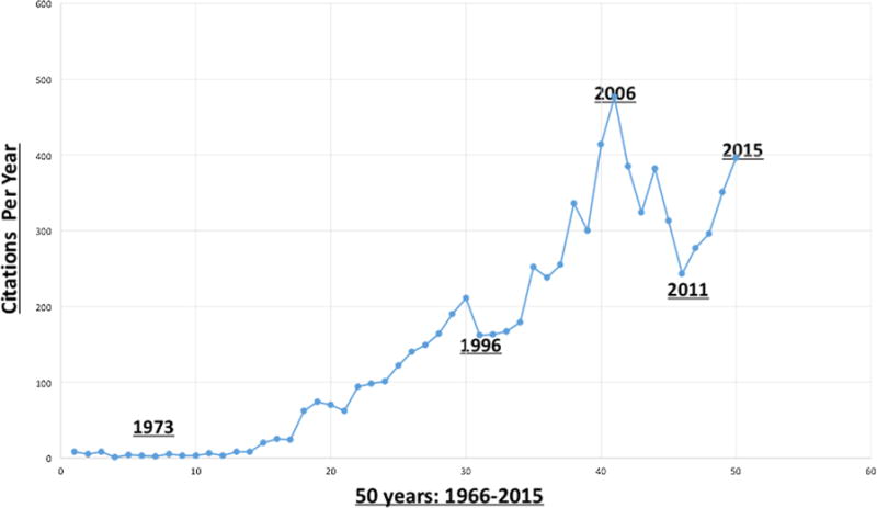
Fifty years’ citations of the key words: “Laser & Gynecology” in peer-review articles (1964– 2015).
The Vagina: Histology and Tissue Properties
The vaginal wall is composed of four layers [18] (Fig. 2): (i) superficial non-keratinized stratified, squamous epithelium; (ii) lamina propria—a dense connective tissue layer; (iii) muscular layer composed of inner circular and outer longitudinal smooth muscle fibers; and (iv) the adventitia—a loose connective, collagen-, and elastic fiber-rich tissue layer, which supports the vaginal wall.
Fig. 2.

Histological sections of the vaginal wall. (A) Normal. (B) Moderately atrophic. (C) Severely atrophic. (A) Estrogenized vaginal histology. The two upper layers of the vaginal wall are shown: stratified, squamous epithelium (SSE), and the lamina propria (LP). The stratified squamous epithelium is rich in glycogen (larger cells with abundant clear cytoplasm—blue arrow) and is nonkeratinizing. The basal cell layer (black arrow) consists of a single layer of columnar cells. (B) Moderately atrophied vagina: Atrophy is shown by thinner epithelium (E) and loss of maturation (smaller cell size with less cytoplasm) on the surface. (C) Marked vaginal atrophy. (Courtesy: Lev-Sagie A).
The mucosal epithelium displays estrogen-dependent behavior and function which naturally reacts to hormonal fluctuations occurring over a woman’s lifetime, as well as during the menstrual cycle [19]. The estrogenized epithelium is rich in glycogen, which is fermented by lactobacilli, lowering the vaginal pH. The lamina propria is primarily composed of collagen and elastin fibers and contains a dense plexus of small blood vessels, lymphatic vessels, and nerves. It is denser toward the surface and freer toward the muscular layer. In the anterior vaginal wall, the lamina propria’s papillae are scarce, but in the posterior wall, they are prominent and deep.
Collagen and elastin participate in the control of the biomechanical properties of the vaginal tissue [18]. Collagen fibers are rigid and do not easily distorted while elastin fibers afford tissue elasticity. Therefore, collagen fibers are the major determinants of vaginal wall strength and its mechanical resistance. Multiple collagen subtypes co-polymerize to form mixed fibrils, whose size and impact on the biomechanical strength of the tissue is largely dependent on the proportion of collagen subtypes within the fiber [20,21]. The main collagen subtypes present in the vagina are I, which forms large and strong fibers, III, which forms smaller fibers of lower tensile strength, contributing to the tissue’s elasticity, and V, which forms small fibers of low tensile strength, which are typically located at the core of the fibril [18].
The effects of estrogens on urogenital tissues are mediated through estrogen receptors (ERs) α and β, which are expressed throughout the urogenital tissues, including the vagina, vulva, urethra, and bladder trigone [22]. In a study evaluating the molecular effect of estradiol on the vagina, differentially expressed mRNA transcripts were detected in vaginal biopsies by microarray analysis before versus after estradiol treatment in menopausal women [23]. Over 3,000 estradiol-regulated genes, involved in several signaling pathways that promote vaginal tissue repair and remodeling were identified, via regulation of cell proliferation, differentiation, apoptosis, pathogen defense, barrier function, inflammation, extracellular matrix metabolism, oxidative stress, and neovascularization, were identified. As a dominant regulator of vaginal physiology, estrogen withdrawal is associated with significant anatomic and physiologic changes in the urogenital tissues. The epithelium becomes pale, thin, less elastic, and progressively smoother as rugal folds decrease. Other changes include reduced collagen content and hyalinization, decreased elastin, altered appearance and function of smooth muscle cells, increased density of connective tissue, and fewer blood vessels (Fig. 2). Furthermore, blood flow and secretions diminish, flexibility, and elasticity of the vaginal vault decrease, the tissues become more friable, vaginal flora changes shifting from a lactobacillus-dominant flora to an anaerobic gram-negative rods and gram-positive cocci flora, and pH increases. It is possible that variations in urogenital symptoms experienced by menopausal women represent inter individual variations in vaginal tissue ER expression and in genetic regulation, among other factors.
Genitourinary Syndrome of Menopause
Genitourinary syndrome of menopause (GSM), also known as vulvovaginal atrophy (VVA), urogenital atrophy, or atrophic vaginitis, is a condition that results from decreased estrogen levels in the urogenital tissues. GSM can occur at any time in a woman’s life cycle, although it is most common in postmenopausal women, when it reportedly affects up to 50% of women [24]. GSM is defined as a collection of signs and symptoms involving changes to the vulva, vagina, urethra, and bladder, including, but not limited to, dryness, burning and irritation, poor lubrication, discomfort or pain, impaired sexual function, and urinary symptoms of urgency, dysuria, and recurrent urinary tract infection [22,24]. The diagnosis of GSM is clinical based upon characteristic symptoms, typical signs on physical examination and laboratory tests. Women may present with some or all of the signs and symptoms, which are bothersome and are not attributed to another disorder. Classic vaginal findings include a pale, dry vaginal epithelium that is smooth and shiny with loss of most rugation.
Laboratory evaluations can confirm the diagnosis and are also used to exclude other diagnoses. The vaginal pH generally exceeds 5, as opposed to the normal pH of an estrogenized vagina, which ranges from 3.5 to 5.0. On saline microscopy, the squamous, large superficial cells are replaced by parabasal or intermediate cells, which are smaller and rounder, and have a relatively large nucleus. A vaginal maturation index (VMI) measuring the proportion of parabasal, intermediate, and superficial squamous mature cells can aid in the diagnosis [25]. In general, measuring serum estrogen levels does help make the diagnosis of atrophic vaginitis [26].
In most cases, GSM is a chronic condition and long-term therapy is required to provide symptomatic relief. Symptomatic GSM can range in severity from bothersome to debilitating symproms, and the extent of adverse consequences of urogenital atrophy makes treatment essential in many women. When left untreated, vaginal symptoms usually worsen [22]. Choice of therapy depends on the severity of symptoms, the effectiveness, and safety of therapy for the individual patient, and patient preference. Treatment goals include symptomatic relief, with reduced systemic exposure to estrogen and minimized potential for adverse effects. Recommended first-line approaches [22] include nonhormonal vaginal lubricants and moisturizers, as well as continued sexual activity. Local estrogen therapy is considered effective and well tolerated for the treatment of moderate to severe symptoms, by re-establishing the pre-menopausal vaginal environment, that is, thickened epithelium, increased secretions, restoration of vaginal flora, and reduced pH. Overall, therapy decreases vaginal dryness and relieves urogenital symptoms. The potential use of energy-based devices to address GSM symptoms is encouraging but calls for more solid scientific data, as is detailed in this review, and treatment protocol guidance, as discussed in the last chapter.
LASER AND RADIOFREQUENCY: PHYSICS AND VAGINAL TISSUE INTERACTIONS
Basic Laser Physics: Ultraviolet to Far-Infrared Wavelength Lasers
Lasers, an acronym for “light amplification by stimulated emission of radiation,” operate in the ultraviolet (157–400 nm), visible (400–800 nm), near-infrared (800–3,000 nm), mid-infrared (3,000–30,000 nm), and far-infrared (>30,000 nm) regions of the electromagnetic spectrum [27]. The wavelengths across the electromagnetic spectrum are differentially absorbed by different tissue chromophores, including hemoglobin, melanin, connective tissue, and water [28]. Selective photothermolysis describes the desirable clinical effect of selectively absorbed laser wavelengths by a chromophore in the target tissue [29]. In practice, to maximize absorption and enhance treatment efficacy, the appropriate wavelength is determined by the main chromophore within the target tissue. For example, melanin and hemoglobin readily absorb light in the visible and near-infrared regions. In contrast, in minimally pigmented tissue, wavelengths highly absorbed by water will provide efficient ablation [30]. In addition, the laser pulse duration can be controlled to confine the thermal damage to the target area. This is achieved by administering a pulse of light that is less than or equal to the thermal relaxation time (τr) of the target (time for the temperature of a Gaussian temperature distribution with a width equal to the target’s diameter to decrease by 50% from the temperature immediately after laser exposure) of the target chromophore in the tissue [30]. Various fractionated beam delivery technologies created microzones of tissue injury, separated by intervening areas of untreated skin, which hastens recovery and reduces the incidence of adverse events [31,32].
Radiofrequency Wavelengths
Radiofrequency (RF) devices emit within a frequency range of 3 kHz to 24 GHz the so-called industrial, scientific, and medical user (ISM) bands. Application of RF for medical use generates an electrical field that results is an oscillating electrical current, which, in turn, induces translational motion of charged atoms and molecules and hinders rotation of polar molecules [33]. This molecular motion, largely responsible for the heat capacity, results in a local temperature increase. Water molecule has a large permanent dipole moment, which is randomly oriented in the absence of an applied electric field. In the presence of an electric field, the dipole moments partially orient along the direction of the field, yet, due to the viscosity of water, energy is required to rotate the dipoles resulting in energy transfer into the tissue [33]. The resistance, or impedance, converts electrical current to thermal energy, generating heat relative to the amount of current and exposure time. Consequently, energy is dispersed to three-dimensional volumes of tissue at controlled depths.
The configuration of electrodes in RF devices can be monopolar, bipolar, or tripolar, all of which have been used for cutaneous applications. In a monopolar system, which has been employed for vaginal applications, the electrical current passes through a single electrode in the hand piece to a grounding pad.
Laser-Tissue Interactions: Ablative Lasers: Carbon Dioxide Laser
The CO2 laser emits light at wavelength of a 10,600 nm, which is strongly absorbed by tissue water (absorption coefficient of 800 cm−1) [29]. The penetration depth is dependent upon the water content, independent of melanin and hemoglobin. The CO2 laser has been traditionally used to ablate tissue, and the uterine cervix is a common target for treatment of various lesions such as genital warts (Fig. 3). Differences between devices in the depth of vaporization, crater base carbonization, and thermal coagulation effect are dependent on the amount of energy deposited as a function of time (Fig. 4). At a pulse duration shorter than 1 millisecond, the CO2 laser light penetrates approximately 20–30 μm into tissue and the zone of residual thermal damage can be confined to a 100–150 μm—thick layer of tissue, although thermal coagulation at a depth of up to 1 mm has been reported [29]. The vaporization or boiling point of water at one atmosphere is 100°C; thus, the energy density required to achieve pulsed-laser ablation of skin tissue is approximately 5 J/cm2. During ablation at these parameters, the skin temperature rises to 120–200°C. The beam diameter also plays an important role in the effect on tissue with small diameter beams (100–300 μm) achieving high fluences and rapid vaporization. Thus, if the beam is not moved rapidly across the skin surface, desiccation, charring, and diffusion of heat may occur. Beams of larger diameters (>2 mm) induce non-vaporization heating and increase the risk of deep thermal damage due to the need to apply low energy densities for longer periods of time before achieving visible vaporization. The super-pulsed or scanned CO2 lasers, combining high peak powers with short pulses and rapid movement across the skin surface, evolved in order to precisely control the depth of ablation and degree of thermal damage.
Fig. 3.
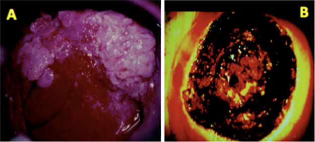
(A) Genital warts (condyloma accuminata) on the uterine cervix. (B) The uterine cervix following superficial ablation of the diseased area. Carbonized crater base is visible. (Courtesy: Levavi H, Tadir Y).
Fig. 4.

Energy (Joule) = Power (Watt)×Time (Second). Effect on tissue is different even if the same energy is deposited depending on exposure time. Same energy may cause different effect, that is, crater shape, superficial carbonization, and thermal coagulation. Three examples of same energy, 90 J, will cause different effect on the tissue. (Courtesy: Tadir Y).
Erbium:YAG Laser (Er:YAG)
The Er:YAG laser is another near-infrared ablative laser used to induce tissue resurfacing. The Er:YAG laser emits light at a wavelength of 2,940 nm, which is close to the absorption peak of water, and yields an absorption coefficient that is 16 times higher than that of the CO2 laser. The Er:YAG laser’s penetration depth is limited to approximately 1–3 μm of tissue per J/cm2, versus the 20–30 μm provided by the CO2 laser [29]. This feature allows for more precise skin ablation, with minimal thermal damage to the surrounding tissues. The estimated resonant-tunneling structure is 10–40 μm. Use of the Er:YAG laser at an energy density of 5 J/cm2 will vaporizes the epidermis after four passes, while an energy density in the range of 8–12 J/cm2 requires only two passes. The variable-pulsed Er:YAG laser, with pulse durations ranging from 10 to 50 milliseconds, elicits immediate tissue contraction and a healing rate that is intermediate between the short-pulsed Er:YAG with pulse durations of 250–350 microseconds and the CO2 laser [31]. The Er:YAG laser is associated with a milder post-operative discomfort, erythema and edema, and overall healing times are faster as compared to the CO2 laser [31]. In contrast, CO2 laser treatment is bloodless, due to its ability to photocoagulate blood vessels of diameters smaller than 0.5 mm, whereas bleeding increases with successive passes with the Er:YAG laser.
Fractional Ablative and Non-Ablative Lasers
In fractional laser resurfacing, an array of microbeams of laser light is delivered to create microscopic columns of energy-mediated effects [31]. The microscopic lesions extend from the epidermal layer into the dermis, or from the vaginal epithelium into the lamina propria, to depths dictated by several parameters, including laser energy density and spot size. The vast majority of fractional ablative devices use an optical scanner to deliver a very small laser spot across the skin. Others employ a “pixelated” technology where an array of laser spots is “stamped” onto the target tissue hough a micro-lens array or holographic beam splitter. The wavelength delivered by all the CO2 laser devices is the same and the diameter of each fractionated microbeam (termed a microspot, or pixel) can range from less than 100 μm to 1.25 mm, depending upon the power density of the beams differing accordingly. The penetration depth of the microbeams varies from less than 50 μm to as deep as 1.6 mm, depending upon the device. Among the fractional Er:YAG laser devices, the microspot size varies from 50 μm to 1.5 mm, and the effect on tissue varies accordingly. An additional technological feature of some fractional Er:YAG devices is a thermal coagulative pulse that may be administered immediately following the ablative pulse to provide additional hemostasis.
Radiofrequency (RF)
In RF-tissue interactions, heat generated in the dermis reaches a thermal dose threshold, above which collagen begins to denature (~60°C) and to fully denature (70–75°C) [34]. Partial denaturation of collagen by RF is maximal at 67°C, and correlates with optimal neocollagenesis and clinical effects in the skin. Temperature at 40–45°C induce production of collagen by fibroblasts and is effective in skin tightening [35]. However, surface temperatures of the skin exceeding 45°C have been correlated with pain and thermal burns during and after RF treatment [36]. No thermal burns have been reported in vaginal tissue treated with RF up to 47°C, however, burns and blisters have been seen at approximately 55°C. Temperature is controlled by using a dedicated software (Alinsod R. Personal communication).
Introduction of mobile RF delivery, where energy is repeatedly delivered within nanoseconds over the same surface area, enabled the cooling of pain afferents at the epidermal-dermal junction, while allowing efficient heating of dermal collagen [36]. Thus, mobile RF delivery, allows for cooling of cutaneous nerves, avoiding a surface temperature exceeding the trigger threshold of 45°C, whereas heat accumulates in the target collagen and other dermal structures, which have a very long τr (225 microseconds) [36]. Current monopolar RF devices for vaginal treatment employ mobile delivery, with target surface temperatures at or below 45°C.
Histological Effects
When applying application of cutaneous energy-based technologies to vaginal wall indications, histological differences between the skin and vaginal mucosa must be considered.
Standard Ablative Skin Histology
Histologic changes observed following ablative skin resurfacing with CO2 and Er:YAG lasers include neocollagenesis approximately 6 weeks post-treatment [28]. Recent reverse-transcriptase polymerase chain reaction and immunohistochemical evaluations from the facial skin of 28 patients following CO2 laser-elicited resurfacing demonstrated upregulation of procollagens I and III, interleukin 1-β, TNF-alpha, TGF-β 1, metalloproteinases (MMP) [27,28,33].
Fractional Ablative and Non-Ablative Effects and Histology
Histological evaluations following fractional CO2 laser resurfacing have demonstrated the direct correlation between energy density and ablative depth of penetration. Increasing the energy density of a fractional CO2 device focused on 135 μm diameter microspot, from 10 to 75 J/cm2 increased the ablative penetration depth from 100 μm to 1.6 mm [31]. The ensuing wound healing process showed granulation tissue at 1–3 days post-treatment, followed by progressive neocollagenesis and dermal remodeling up to 30 days post-treatment. Neocollagenesis continued for several months thereafter, as has been observed following standard ablative CO2 laser resurfacing [31].
Assessments of the effects of the fractional CO2 ablative laser on the vaginal mucosa demonstrated an increase in squamous stratified epithelium thickness, with augmented levels of glycogen in the epithelial cells and a large number of glycogen-rich cells being shedded at the epithelial surface [14,15]. Active fibroblasts were detected in the connective tissue of the lamina propria, as well as increased extracellular matrix content, including collagen, and ground substance. In addition, newly formed connective tissue papillae, undulated epithelium, and typical blood capillaries penetrating inside the papillae were observed post-treatment [14,15]. Similarly, fractional Er: YAG laser treatment of vaginal mucosa led to increased thickness and cellularity of the epithelium and a more compact lamina propria with a denser arrangement of connective tissue. Increases in collagen and elastin content were also demonstrated [37].
Unlike the CO2 laser, which causes superficial vaporization and deeper thermal effects, the Er:YAG laser is a true ablative laser. The Er:YAG laser only produces a thermal effect at depths of about 5–20 μm per impact as opposed to the 50–125 μm-deep incremental thermal effect with each pass of the CO2 laser. Use of the Er:YAG laser with minimal ablation reportedly resulted in vaginal rejuvenation and improved control of urination [38]. Attempts to use the same 2,940 nm laser in a non-ablative (Smooth) mode resulted in similar effects. The “smooth mode pulse,” with a duration of 250 milliseconds, consisted of a fast sequence of individual super-pulse mode (300 microseconds) micropulses with intra-pulse intervals of 50 milliseconds [39]. In this mode, vaginal mucosa temperatures increased to 60–65°C, without inducing superficial ablation.
RF energy is dispersed in three-dimensional volumes of tissue at controlled depths. The creation of new dermal volume in response to RF treatment has been extensively reported, and has been shown to improve skin laxity and the skin mechanical characteristics of the skin [40]. Both neocollagenesis and elastogenesis are induced with improved skin elasticity, which correlates with elastometry [40]. Collagen fibers are composed of a triple helix of protein chains linked via interchain bonds to form a highly organized structure. When collagen fibers are heated to specific temperatures by any energy based devices, they contract due to breakage of intramolecular hydrogen bonds. Contraction causes the triple helix structure to fold, creating thicker and shorter collagen fibers which are thought to be the mechanism of action of immediate tissue tightening seen after skin-resurfacing procedures. The partially denatured collagen serves as a signal for neocollagenesis [34]. The creation of new elastin, which is relatively unique to RF, may play a role in its effectiveness in treating vaginal laxity [34,40]. Histological studies of vaginal mucosa will be necessary to evaluate the effect of monopolar RF on symptomatic relief of GSM and on vulvovaginal prolapse. Cell activation and tissue rejuvenation is discussed in the next section.
The Technology
Beam splitting into multiple microbeams can be achieved by rapid computer controlled scanning, or with a holographic beam splitter, which prints pixelated microbeams on the target tissue and can be used in both ablative and non-ablative mode in order to induce cell activation (Fig. 5). These changes allowed the conversion of energy-based devices from cutting and coagulating tools to cell activation and tissue rejuvenation platforms. Lessons learned of the effect of such devices on skin cells, have been translated to the vaginal mucosa in an effort to reverse the natural process of aging (Fig. 6).
Fig. 5.
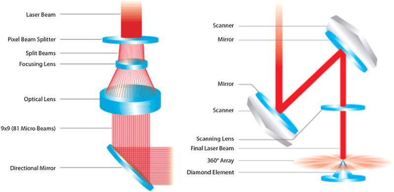
(Left) Laser beam fractionated by holographic lens, printing 9×9 (81 microbeams) on 10 mm2. (Right) Laser beam delivered through computer controlled, two parallel mirrors and scanning lens. (Courtesy: Alma Lasers).
Fig. 6.
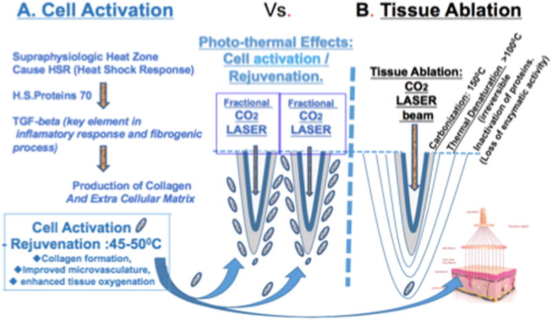
(A) Fractional micro-ablation inducing cell activation and tissue rejuvenation at 45–50°C [42]. (B) Tissue ablation and thermal effects on adjacent layers (Courtesy: Tadir Y).
Fractional laser technology, which creates a pattern of microscopic ablative zones, surrounded by adjacent normal skin, was a milestone in the history of light-based tissue rejuvenation. Suprathreshold energy density of fully ablative therapy results in cellular vaporization, and subablative fluence induces irreversible coagulation and protein denaturation through heat transfer [41]. Thermomechanical tissue destruction (ablation) driven by light based devices, generally extends 200–300 μm deep in the tissue and is followed by tissue tightening through the process of heat-induced shrinkage of collagen and neocollagenesis. This occurs at temperatures between 45 and 50°C in the in the zone surrounding the ablated tissue. As part of the wound healing process, live cells react to this temperature increase with a heat shock response (HSR), which can be defined as a temporary change in cellular metabolism characterized by the production of a small family of proteins defined as heat shock proteins (HSP). Studies on the skin have demonstrated that HSP 70 plays a role in the coordinated expression of growth factors such as transforming TGF-β, which is known to be a key element of the inflammatory response, fibrogenic process, and in the production of new collagen and extracellular matrix [42]. A collection of vaginal and vulvar probes is presented in Figure 7, listed in alphabetic order and according to the type of energy source.
Fig. 7.
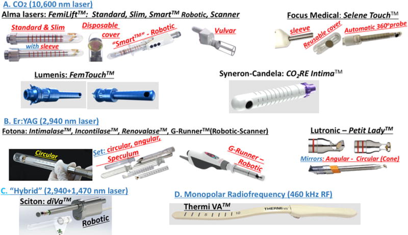
Energy-based probes for vaginal and vulvar treatment listed in alphabetic order. (Courtesy: manufacturers).
The width of the presented vaginal laser probes varies between 19 and 38 mm, and the RF probe is 15 mm, all designed to accommodate the pre- and post-menopausal vagina demensions. Several laser probes integrate fractional/pixel stamping technology (Alma Lasers, Buffalo Grove, IL; Focus Medical, Bethel, CT; Fotona, San Clemente, CA) and some are based on tissue scanning technology (Focus, Lumenis, San Jose, CA; Syneron-Candela, Irvine, CA; Fotona, and Sciton, Inc., Palo Alto, CA). Most delivery systems are operated manually at 90° angle; some offer 360° circular exposure (Fotona, Focus, Lutronic), and some are fully automatic, robotic-computer-controlled probes (Alma’s FemiLift Smart™, Fotona’s G-Runner™, Sciton’s diVa™”). ThermiVa (Irving, TX) RF probe is designed to treat the vagina and vulva.
Unlike Er:YAG and several other laser wavelengths, the commonly used CO2 laser beam does not pass through the disposable plastic cover of a reusable vaginal probe. As such, a biocompatible lens located at the beam path of the sterile cover allows use of a disposable, sealed sterile hand piece (Alma Lasers, Focus). Some manufacturers offer sterile single-use handpiece (Syneron-Candela) and some offer laser-dedicated vaginal speculum (Fotona, Lutronic). Technical parameters are listed in Table 1.
TABLE 1.
Technical Parameters of Vaginal Probes Listed in Manufacturer’s Alphabetic Order (Courtesy: Manufacturers)
| Brand name | Laser type/wavelength (nm) or RF | Pulse duration (ms) | Maximum energy/pulse (mJ) | Surface area “lased”/exposure (mm2) |
|---|---|---|---|---|
| Alma Lasers, Buffalo Grove, IL. | CO2 – 10,600 | 400 | 500 (per pixel) | 10 |
| Fotona, San Clemente, CA. | Er:YAG – 2,940 | 250 | 240 J (per pass) 3 J/cm2 |
80 (cm2), non-ablative, entire surface |
| Focus Medical, Bethel, CT. | CO2 – 10,600 | 1–200 | 60 | 10 |
| Lumenis, San Jose, CA. | CO2 – 10,600 | NA | 7.5/10/12.5 | NA |
| Lutronic Aesthetics, Burlington, MA. | Er:YAG – 2,940 | 0.2 ~ max. 1500 (Dual mode) | 3.7 J (per pulse) | 144 (per pulse) |
| Sciton, Inc. Palo Alto, CA. | Hybrid: 2,940/1,470 | 150/20 | 300/100 | 1.5–2.5 |
| Syneron-Candela, Irvine, CA. | CO2 – 10,600 | 20–1,066 | 70 | 10 |
| ThermiVA, Irving, TX. | RF 460 kHz | – | Estimated tissue temp. 47°C | 10 |
NA: not available.
ABLATIVE LASER-INDUCED CHANGES IN THE VAGINAL WALL
Technical parameters of energy-based devices and the distinct physiological characteristics of the target tissue, directly influence the cellular activations driving tissue restoration. These include laser wavelength, energy density, pulse duration, spot diameter, tissue absorption and hydration, tissue oxygenation, blood supply, degree of keratinization (in the case of skin), and insulating properties (adipose tissue). The energetic surcharge endured by an excessively high increase in tissue temperature can jeopardize cell survival. In the case of vaginal mucosa, in which epithelial cells and the ground substance of the connective tissue are rich in water, it is important to consider that healthy premenopausal mucosa is highly hydrated while the atrophic postmenopausal mucosa is characteristically dry [43]. Therefore, a controlled power of the source must be used.
Based on successful fractional CO2 laser treatment of aging-related skin changes [44,45], and on preliminary experience with the fractional CO2 laser applied for vaginal rejuvenation [14], the irradiation parameters of fractional CO2 laser were further optimized through an ex vivo study on the vaginal mucosa as MonaLisa Touch™ (DEKA, Florence, Italy) treatment [14]. A pilot study on 50 postmenopausal women suffering from severe symptoms of vaginal atrophy demonstrated the effectiveness of this treatment [46]. Clinical efficacy, including breast cancer survivors, for whom estrogen treatment is contraindicated, was confirmed by various authors [47–51]. Histologically evident modifications of the postmenopausal atrophic vaginal mucosa following fractional CO2 laser treatment has provided a structural basis for understanding the molecular mechanisms responsible for restoration to a healthy mucosa [14,15,46].
REJUVENATION OF THE VAGINAL MUCOSA
Premenopause
As mentioned earlier, the premenopausal vaginal mucosa is characterized by a squamous stratified non–keratinized epithelium and a lamina propria of connective tissue, often protruding into the undersurface of the epithelium, with papillae rich in small blood vessels. Considering the absence of glands in the vaginal mucosa, epithelial cell shedding represents a sort of secretory activity critical for vaginal health. Starting from the basal layer, where epithelial cells proliferate in order to compensate for the loss of the cells shedded at the mucosal surface, epithelial cells undergo a process of differentiation. Glycogen begins to be synthesized by the cells of the intermediate layers and is subsequently stored in the superficial cells. When the most superficial cells are shed into the vaginal lumen, their glycogen content is deposited at the epithelial surface, forming favorable conditions for Lactobacilli activity, which is a key factor in maintaining the low pH of the healthy inner vaginal environment.
The connective tissue is rich in blood vessels, which penetrate as small capillaries inside the papillae, providing metabolic support (nutrients, oxygen, and other molecules) to the intermediate and superficial cell layers. Collagen fibers are organized in compact bundles of different orientation which, together with elastic fibers, constitute an effective support for the mucosal part of the vaginal wall. The ground substance, comprised of glycosaminoglycans, proteoglycans, and multiadhesive glycoproteins, is a significant component of the extracellular matrix of the connective tissue and represents the most permeable part of the matrix. Its components are particularly rich in polar groups capable of linking large quantities of water molecules and are responsible for the high hydration status (turgidity) of the mucosa. The consequently high permeability of the matrix permits easy diffusion of water, ions, nutrients, signaling molecules, and many other molecular species through the extracellular matrix.
Postmenopause
The decline and later arrest of ovarian estrogen production correlates with significant structural and functional changes in the vaginal mucosa, resulting in atrophy, characterized by significant thinning of the epithelium with a reduction of epithelial renewal dynamics and the directly related absence of superficial cell desquamation. The epithelium-connective tissue interface appears smooth due to the reduction and/or absence of papillae and blood vessels (Fig. 8A). In the connective tissue, fibroblasts are characterized by a small cytoplasm around the nucleus and by frequent projections of very thin processes into the surrounding matrix. In addition, cells exhibit a lower number of organelles, particularly of the rough endoplasmic reticulum and the Golgi apparatus, both involved in the synthesis and turnover of the molecular components of the extracellular matrix, functions significantly reduced in the postmenopause.
Fig. 8.

Trichrome staining of biopsies from atrophic vaginal mucosa before (A) and vaginal mucosa following the fractional CO2 treatment 2 months from the first laser application (B). Particularly appreciable are the remarkable differences: (A) thin epithelium, with small size and few layers of epithelial cells, no superficial desquamation, smooth surface of the interface epithelium-connective tissue, absence of papillae, compactness of the connective tissue. (B) Very thick epithelium, with many layers of big cells, many shedding cells at the epithelial surface, uneven interface epithelium-connective tissue, a well visible deep papilla formed by a fine fibrillar connective tissue with vessels inside (A and B same magnification). Due to the thickness of epithelium following treatment, Figure 8b is formed by two frames. (Courtesy: Calligaro A).
POSTMENOPAUSAL MUCOSA AFTER FRACTIONAL CO2 LASER TREATMENT
The post-treatment stratified squamous epithelium is thicker and comprised of 20–40 cell layers which provide cells for differentiation and superficial shedding. Basal layer cells appear closely packed as in continuously renewing stratified epithelium. The intermediate layer cells appear enlarged, with the nucleus surrounded by a wide cytoplasm. In the most superficial layer, many wide cells, shedding from the epithelial surface into the vaginal lumen, are observable (Fig. 8B).
The basal surface of the epithelium appears characteristically indented due to the presence of numerous papillae of connective tissue, protruding onto the undersurface of the epithelium, yielding an uneven appearance to the epithelial-connective tissue junction. Elongated blood capillaries inside the papillae are also clearly observable (Fig. 9). Reliable histochemical methods such as Periodic acid-Schiff (PAS) and Alcian blue staining, enabled detection of large quantities of glycogen in the cytoplasm of epithelial cells of the intermediate and superficial layers, and inside the numerous exfoliating cells of the most superficial layer. The comparison with the atrophic mucosa before treatment is presented as Figure 10.
Fig. 9.

Common feature following treatment. At the center of the figure, a papilla with a small and long vessel inside is easily identifiable due to its erythrocyte content and the thin endothelial profiles. In the insert, numerous newly formed papillae following treatment are recognizable. H&E staining. (Courtesy: Calligaro A).
Fig. 10.

PAS staining of vaginal mucosa sections from biopsies before (A) and following treatment (B). Besides the evident difference in the epithelial thickness, the PAS signal shows (A) few small cells containing glycogen (fuchsin-stained) versus (B) many larger cells stained, starting from the low intermediate cell layer. Also of note in (B) is the superficial desquamation of intensely stained cells, which deposit glycogen at the surface of mucosal epithelium. (Courtesy: Calligaro A).
Epithelial changes induced by the laser treatment, and the renewed cells differentiation with synthesis, storage, and delivery of glycogen (Fig. 11), are the consequence of the delivery of cytokines/growth factors in the connective tissue stimulating a renewal of its structure and functionality. This is achieved by a full restoration of the connective tissue, both fibers (mainly collagen) and the ground substance, rich in highly hydrophilic molecules. In this way, the hydration and permeability of the extracellular matrix are improved, permitting a high rate of molecular traffic both inside the connective tissue and between the newly formed vessels and the epithelium, with restoration of related epithelial functions (proliferation, differentiation with a renewed glycogen synthesis, and desquamation).
Fig. 11.
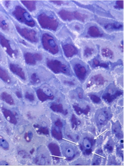
High-resolution, light microscope image of a CO2 laser treated-section, embedded in epoxy resin for electron microscopy, and stained with toluidine blue. In the lower part of the image, some cells of the lower intermediate layer with a very clear nucleus containing a highly visible nucleolus (as a dark blue spot), show some violet-stained masses (white arrows). These represent the first stores of newly synthesized glycogen inside epithelial cells, resulting from the restored differentiation process. In the upper layers, glycogen masses are increasingly extended to almost completely fill the cytoplasm, until superficial shedding (desquamation). (Courtesy: Calligaro A).
Comparative microscopic and ultrastructural analyses of biopsies collected before versus after fractional CO2 laser treatment, demonstrate the recovery of all the mucosal structures, suggesting restoration of full physiological functioning, both in the epithelial and connective tissue compartments. The observed modifications of the mucosal structures following fractional CO2 treatment are summarized below (Figs. 8–12).
Fig. 12.

Electron microscopy of fibroblasts in samples collected following fractional CO2 treatment. (A) Connective tissue fibroblasts showing a relatively compacted cell body, with a cytoplasm appearing almost completely filled with rough endoplasmic reticulum (rER). The nucleus is euchromatic, with a well-presented nucleolus. These features support active synthesis of proteins (collagen and others) to be delivered to the surrounding extracellular matrix, promoting its renewal. (B) In fibroblasts, close to the rER profiles (cisternae formed by membranes with ribosomes attached at their surface), dilated cisternae with attached ribosomes are frequently observed as vesicles containing a fine, filamentous material, representing the molecular precursors of extracellular fibrillar components. (C) A Golgi apparatus is clearly visible. Two Golgi complexes, formed by stacks of membranes with associated dilations and vesicles, are recognizable (white arrows). (Courtesy: Calligaro A).
Epithelium
Following the fractional CO2 laser treatment, the epithelial structure is fully restored, due to newly active proliferative and differentiative processes of epithelial cells, with thickening and desquamation. The lactic acid-producing Lactobacilli vaginalis (bacteria physiologically resident in the vagina) feed on glucose derived from the glycogen delivered by shedding epithelial cells, acidify the vaginal transudate fluid at the mucosal surface, and thereby restore the vaginal environment to the acidic premenopausal state, and prevent the colonization of yeast and other bacterial pathogens [52,53].
Connective tissue, lamina propria fibroblasts are characterized by high organelle content, an extended rough endoplasmic reticulum, which is the site of procollagen synthesis [54], and frequently dilated cisternae (Fig. 12A and B). A well-developed Golgi apparatus with flattened stack of membranes and associated vesicles (Fig. 12C), the site of synthesis of polysaccharides and of the glycosylation of the protein components of the ground substance, such as glycoproteins, proteoglycans, and multiadhesive glycoproteins, is also detectable [55]. These structural features provide the machinery necessary for the synthesis of both the fibrillar and the ground substance components of the extracellular matrix. In parallel, the high blood vessel content in the connective tissue supports the renewed activity of fibroblasts and capillaries penetrating the newly formed papillae underneath the epithelium (Fig. 9), and provides metabolic support for epithelial cell proliferation and differentiation.
Significant amounts of data relating to the impact of CO2 laser treatment on human skin, were obtained using reverse transcriptase real-time polymerase chain reaction (PCR) technology and immunohistochemistry [56]. The reported highly significant increase in the mRNA levels of procollagen, metalloproteinases (MMPs), primary interleukin-1β, tumor necrosis factor-α, and profibrotic cytokine TGF-β1 following CO2 laser application to the skin suggests stimulation of tissue renewal mechanisms, which seemingly occurs in the vaginal mucosa as well. Stimulation of connective tissue matrix remodeling by fibroblasts following fractional CO2 laser application has been tightly correlated with the activation of heat shock proteins (HSPs). Immunohistochemical analysis of HSP in human fractional CO2 laser treated skin revealed a persistent collagen-associated response persisting at least 3 months following treatment [57]. The collagen chaperone HSP47 [58,59] localized in the rough endoplasmic reticulum plays an important role in the early stages of post-treatment collagen biosynthesis [60]. In the application of fractional CO2 laser on human skin [61], the local increase in different cytokines has been histochemically demonstrated, particularly transforming growth factor-β (TGF-β; stimulating matrix proteins, such as collagen), basic fibroblast growth factor (bFGF; stimulating angiogenetic activity with endothelial cell migration and proliferation), epidermal growth factor (EGF; stimulating the re-epithelization), platelet-derived growth factor (PDGF; stimulating fibroblasts to produce extracellular matrix components), and vascular endothelial growth factor (VEGF; regulating vasculogenesis and angiogenesis) (Fig. 6A). These observations [61] closely parallel the observed microscopic and ultrastructural modifications of the postmenopausal atrophic vaginal mucosa following fractional CO2 laser treatment, that is, regulated stimulation of fibroblast-driven renewal of the extracellular matrix and activation of epithelial cell differentiation. The expression of these mechanisms is initiated in the first suprabasal layers of the epithelium (glycogen synthesis) and in the connective tissue with the stimulation of fibroblast activity and the formation of new vessels. Other studies on human skin corroborate these findings relating to increased cytokines/growth factor expression following fractional CO2 laser application [62,63].
Deployment of the fractional laser for the treatment of the atrophic vaginal mucosa leads to neocollagenesis and to production of ground substance components within the connective tissue, and glycogen and acidic mucins within the epithelium and on the epithelial surface. These activities revert the vaginal mucosa from an atrophic state to a healthy premenopausal state, resulting in highly significant symptomatic relief [46].
Sub-ablative (or non-ablative) long pulse modes have been evaluated for their ability to induce deep thermal effects on the surface of the vaginal mucosa. Deep collagen coagulation by stacking repetitive Er:YAG laser pulses on the same tissue site has been described initially by numeric modeling [64]. This mode of laser application on the mucosa can generate a thermal change in deeper layers, with only minor superficial ablation (5 μm) on the surface, but with sufficient caloric effect to thermally alter the chromophore. Using a large spot size (>5 mm), the heating process can be achieved due to the permanence of the long-pulse stimulus and the repetition of exposures. This deepens the heat effect in the lamina propria to a depth of at least 500–1,000 μm (depending on the degree of tissue hydration), inflicting the known “Joule effect” (photothermal and thermochemical effect) on the lamina propria of the vaginal mucosa. In turn, a local rise in temperature, release of bradykinin and histamine with relaxation of precapillary arteriolar sphincters, and vasodilation are achieved in an overall effect called “The Phenomenon of Thermal Reperfusion” (FTR). Deep coagulation of collagen by repetitive Er:YAG laser pulses has been predicted by theoretical models, demonstrated on animal skin [65,66], and confirmed in the vaginal wall [67].
These technologies open new perspectives on the potential use of electromagnetic radiation to re-establish structural and physiological conditions in other genitourinary anatomic sites. Other non-laser energy sources such as RF and High Intensity Focused Ultrasound (HiFU) are being tested for vaginal rejuvenation as well.
Vaginal Tightening focuses on restoring functionality by reversing the deterioration of vaginal wall structures resulting from natural aging and physical trauma accompanying childbirth. Lack of strength and support of vaginal tissues correlates with reduced sexual pleasure, as first described in the 1960s by Masters and Johnson [68]. Surgical techniques for vaginal tightening aimed to modify the diameter of the vaginal canal by surgically removing extra tissue. In severe cases, the approach is more invasive and targets the fascia approximation and muscular plication. Microablative fractional lasers [13], and RF [69–71] devices induce superficial tissue shrinkage as well as deep stimulation of the collagen layer of the submucosa. Unlike skin complications such as hypertrophic scarring, ectropion formation, and disseminated infection, which have all been reported following fractional laser skin resurfacing [72]. No major complications have been reported following energy-based vaginal rejuvenation procedures.
Urinary Incontinence (UI)
Involuntary leakage of urine is called urinary incontinence, and stress urinary incontinence (SUI) is the most prevalent type caused by transient increase of intra-abdominal pressure and weakening of support naturally provided by the bladder, urethra, and pelvic structures. In healthy states, this support is primarily provided by collagen and elastic connective tissue which degenerate with age and under various pathological conditions [73]. Urethral support can be improved by exploiting the energy-based tightening effect. Numerous publications confirm enhanced urinary control following treatment of SUI with an Er:YAG laser [74–78], CO2 laser [79], and RF [80] all of which triggers a photo-thermal effect, as deep as 0.5 mm inside the vaginal wall, and results in a 30% reduction in tissue volume. Mechanical pull by the deeper tissue layers following shrinkage of the upper, photothermally processed tissue layers, further contributes to the tightening effect. At the same time, thermally induced neo-collagenesis improves thickness, elasticity, and firmness of the vaginal wall [75,76].
Er:YAG laser treatment for SUI has been performed with an accessory kit, (Fig. 7B, Incontilase™, Fotona), composed of two hand pieces (full spot and fractional or patterned spot), two adapters or extensions (angular and circular) to deliver energy within the vaginal canal, and a laser speculum, which facilitates opening of the vaginal canal and serves as a guide for the handpiece adapter. Using the circular adapter and full spot handpiece, total energy of about 650 J is delivered along the entire length of the vaginal wall. In the second phase, the patterned spot handpiece is attached to the angular adapter to irradiate (250 J) the anterior, sub-urethral vaginal wall. The fractional handpiece, delivering energy (~100 J) to the introitus area, is then deployed to repair connective tissue around the urethra and to shrink the introitus. All three phases are done in one treatment session. This laser system effectively addressed various grades of SUI and overall urinary control by triggering a rejuvenation effect in various structures of the mucosa, that is, the epithelium and the lamina propria [76–78], with optimal results achieved following three treatment sessions performed at 4-week intervals. Statistically significant improvements were noted in incontinence severity indices (ISI) and in the degree of incontinence. In the largest published study of 175 women treated with this protocol, 77% of the SUI patients were dry, while only 34% of the patients presenting with a combination of both, SUI and urge incontinence (mixed UI), remained dry at 1-year follow-up. At 2 years post-treatment, 82.8% of the patients in the group with improved outcome remained dry, while 17.2% of patients developed mild incontinence. Three years after the initial treatment, the regimen was repeated in 25% of patients to address mild incontinence. No difference in treatment efficacy was noted between pre- versus post-menopausal patients [75].
In another study (N = 35) in which same protocol is repeated for the treatment of SUI, urodynamic studies, pad testing, lower urinary tract symptoms (LUTS), and sexual function questionnaires were assessed before and after treatment. Among 28 women with baseline pad weights >1 g, 11 (39.3%) were objectively cured and 11 (39.3%) improved. Among the 18 women with mild SUI (i.e., pad weight 1–10 g) nine (50%) were cured and five (27.8%) improved. Among 32 women with complete questionnaire data at 6 months, seven (21.9%) were subjectively cured, and four (12.5%) improved. The authors concluded that the procedure for mild SUI was moderate at 6-month follow-up, but was not effective for pad weight >10 g. Moreover, it improved LUTS, QoL, and sexual function [78].
A prospective observational study of the efficacy of fractional CO2 laser in post-menopausal women with moderate-severe clinical signs (N= 53) showed significant improvement of dyspareunia, dryness, burning, itching, dysuria, frequency, urgency, urgency, and SUI scores assessed by various standard questionnaires. Participants received intravaginal therapy, once a month for 3 consecutive months, with a CO2 laser system (MonaLisa Touch™, DEKA, Florence, Italy) and the setting were performed as previously described [79]. Urinary symptoms improved significantly, as reflected by the significant reduction in the scores of the relevant urinary and quality of life questionnaires. Over the course of the study, the number of patients with lower urinary tract symptoms was improved significantly as reflected by various scores. All participants showed a >5-point improvements in the King’s Health Questionnaire (KHQ) score, which includes objective assessments of symptoms and strong psychometric aspects of urinary incontinence. Factors predictive of ideal CO2 laser therapy candidates were not identified [79].
Non-ablative monopolar transcutaneous temperature-controlled radiofrequency (TTCRF) has been tested for its efficacy in treating SUI symptoms in a small group women (N = 10/cohort) and compared to sham treatment [80]. The vaginal sidewalls, as well as the anterior and posterior walls, were heated to a temperature of 40–45°C by sweeping “half-moon motions” of the RF probe (Fig. 7D, RF). Treatment time was 3–5 minutes per zone. TCRF treatment was associated with a significant (P < 0.01) improvement of ICIQ-SF and UDI-6 scores. Seven of 10 patients (70%) had a negative cough stress test after treatment; improvements were maintained for up to 12 weeks. The clinical outcomes were supported by the positive histologically detected vaginal changes seen in women suffering from postmenopausal vaginal atrophy. TTCRF was well tolerated with no complications reported by treated patients. While the cohort size in some of these studies was small, and duration of follow-up short the results are encouraging.
EXTERNAL GENITALS
Anatomy and Histology
The skin of the external genitals is comprised of the epidermis (E), the dermis (D), and the subcutaneous tissue (SG) (Fig. 13). The epidermis consists of a stratified squamous epithelium, containing basal cells (which divide to form keratinocytes), and keratinocytes. Cells divide in the basal layer and move up, changing their appearance as they move from one layer to the next, resulting in the continuous replacement of cells in the epidermal layer. The keratinocytes synthesize insoluble protein which remains in the cell and later becomes a major component of the outer layer, called the stratum corneum. The dermis is composed of three types of connective tissue: collagen, elastic tissue, and reticular fibers. It is divided into an upper, thin papillary layer, which is composed of thin randomly arranged collagen fibers, and a deeper reticular layer, composed of thick collagen fibers arranged parallel to the surface of the skin surface [81].
Fig. 13.
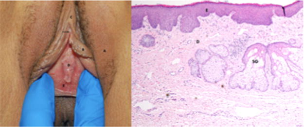
Normal vulva and histology. Left: Hair bearing vulvar skin A-labia majora, B-labia minora, C-clitoral hood, D-Hymen, E-vestibule. black arrow-clitoris, red arrow-Hart’s line. Right: Histology of the non-hair bearing vulvar skin. This is hair bearing skin, ×40 magnification. The epidermis (E) does not have estrogen receptors, so does not atrophy like the vagina when estrogen is withdrawn. D-dermis. There are superficial sebaceous glands (Fordyce spots) present (SG). Like hair bearing skin, the labia minora atrophy when estrogen is withdrawn because the dermal fibrioblasts have estrogen receptors. (Courtesy: Lev-Sagie A).
The vulva exhibits several different types of epithelium [82]. The lateral aspect of the labia majora is covered with dry, keratinized, hair-bearing skin. The medial aspect of the labia majora and the entire labia minora display moist, modified mucous membrane, comprised of a partially keratinized epithelium that contains subtle hair follicles, apocrine sweat glands, and sebaceous glands. A variably distinct line of demarcation termed the “Hart’s line” is evident at the base of the medial aspect of each labium minus, separating the modified mucous membranes of the labia minora from the mucous membrane of the vestibule [82]. This line also demarcates the boundary between the embryonic ectoderm (labia minora) and endoderm (the vestibule). The tissue medial to the Hart’s line is an unkeratinized, non-hair-bearing epithelium with mucous secreting glands. The hymen separates the vestibule from the vagina (Fig. 13).
Lichen Sclerosus (LS) is a chronic, benign, inflammatory skin disease of unknown etiology which can occur at any site but has a predilection for the genital area and rarely involves the vagina. In the majority of patients, the disease is progressive, causing vulvar scarring, loss of portions or all of the labia minora (resorption), clitoral adhesion and narrowing of the introitus [83] (Fig. 14A and B).
Fig. 14.
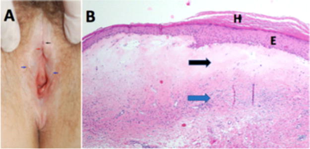
Vulvar lichen sclerosus—(A) LS can be diagnosed clinically by inspection of the vulva for the characteristic thin, white, wrinkled skin, and changes in vulvar architecture. Findings in LS include hypopigmentation, hemorrhages (black arrow), loss of normal architecture including disappearance of labia minora (blue arrow), buried clitoris (red arrow), and narrowing of the introital opening. Note that the distinction between the labia majora and minora is lost. The disease involves the perineal and perianal areas. (B) LS histology ×40 magnification—on histological examination, the epidermis (E) is typically thinned (accounting for the older nomenclature “lichen sclerosus et atrophicus”), although areas of thickened skin (H-hyperkeratosis) may exist. The upper dermis exhibits homogenization of collagen (black arrow) with a band of lymphocytes below this region (blue arrow). (Courtesy: Lev-Sagie A).
Vulvar LS can occur at any age, and its true prevalence is not known, but is estimated at 1 in 59 women in a general gynecology practice [84]. The causes for LS are unknown, but several mechanisms have been studied, suggesting multifactorial origin, with involvement of genetic, hormonal, autoimmune, and inflammatory factors. Symptoms include pruritus, soreness, irritation, and pain. Patients may also complain of dyspareunia, dysuria, and peri-anal involvement although uncommon, active disease may be asymptomatic. The diagnosis of vulvar LS is based upon the presence of characteristic clinical manifestations, ideally with histological confirmation [85]. Most frequently, it affects the labia minora and/or labia majora, clitoris and clitoral hood, and it may also involve the perineum and the perianal skin.
LS usually requires permanent management to maintain remission [85]. If left untreated, it has significant potential to result in the progressive destruction of the vulvar architecture and less commonly, has been associated with vulvar squamous cell carcinoma (SCC) [85]. Topical corticosteroids are the mainstay of therapy, and super-potent topical corticosteroids, such as clobetasol propionate, have long been considered the standard of care for vulvar LS. Recommended therapy usually consists of daily application of corticosteroid ointment for 6–12 weeks, followed by maintenance therapy two to three times per week, if symptoms improve [85].
Medical therapy leads to symptomatic relief in most women; however, the incidence of long-term sequelae, such as scarring and SCC, is unclear, although adequate treatment seems to be associated with a reduced risk of development of neoplasia [86].
Superficial ablation of LS by means of a CO2 laser has previously been described [87–89]. The technique requires use of general anesthesia and a healing period of several weeks. The high cost for the laser tools and the need to perform such procedures in surgical facilities has limited widespread embracement by physicians and patients. However, advancements in fractional laser technology, which do not entail use of general anesthesia, and incur minimal superficial ablation alongside thermal cell activation and tissue rejuvenation, have increased the potential use of this treatment approach [90]. Several reports of LS treated successfully with fractional CO2 laser are available (Table 2). In a recent prospective study, symptomatic postmenopausal patients (N= 27) diagnosed with LS were treated with a fractional CO2 laser, three sessions 4–6 weeks intervals [91]. Twenty-four patients reported cessation of itching and pain, and in 26 patients visible improvement of skin color, elasticity, and vascularity was noticed. No histology is available in this study following treatment.
TABLE 2.
Studies Using CO2 Laser for the Treatment of Lichen Sclerosus
| Author and year | Trial design | Primary outcomes | Findings | Complications |
|---|---|---|---|---|
| Kartamaa and Reitamo (1997) [87] | Case series | Subjective assessment of lichen sclerosus (better, asymptomatic, some recurrence) | Among male and female patients (N = 10) with biopsy proven refractory LS, 76% were asymptomatic after CO2 laser treatment with mean follow up of 32 months. | Three patients needed repeat treatment |
| Peterson et al. (2004) [89] | Case report | Subjective symptoms visual re-epithelization | All patients (N= 2) with refractory anogenital lichen sclerosus had resolution of symptoms and re-epithelization after 2–3 years with CO2 laser therapy | None |
| Windahl (2006) [88] | Retrospective cohort | Subjective patient assessment of recurrence of symptoms, any visible penile lesion, recurrence of meatal stenosis | In men with histologically verified penile lichen sclerosus treated with CO2 laser, 80% (N = 50) had no local symptoms at mean follow-up 14 years | Two patients required further treatment |
| Lee et al. (2016) [90] | Case series | Subjective symptom resolution | In women (N= 5) with biopsy confirmed severe vulvar LS recalcitrant to topical corticosteroid treatment; four of whom responded positively to fractional CO2 laser treatment, and one to ablative CO2 laser therapy | Two patients needed repeat treatment at 6–8 months. Two patients reported discomfort with laser treatment |
| Baggish (2016) [91] | Prospective (N = 27) | Subjective symptom resolution | 24/27 cessation of itching and pain. 26/27 visible improvement of skin color, elasticity, and vascularity | None |
Two additional cases with typical symptoms and histologically proven LS who failed long-term conventional treatment with clobetasol 0.5% and 2% testosterone cream underwent successful treatment with fractional CO2 laser. Unlike conventional symptomatic treatment, histological evaluations before and after the laser treatment demonstrate curative tissue changes (Fig. 15. Elias J, personal communication). Three treatment sessions were conducted at 1 month intervals. Three passes were made at each session, with the Alma Laser—pixel hand piece set, on low to medium energy (10–20 mJ/pixel). Patients became asymptomatic shortly after the third treatment session, and the vulva, introitus and clitoral areas appeared healthy, with elastic closure of the introitus. Tissue appearance was maintained throughout the 6 month post-treatment follow-up period. Histological assessment of samples collected 45 days following the last treatment session showed a trophic epithelium with acanthotic areas without superficial hyperkeratosis. Prospective trials that are in progress may determine if this treatment concept is valid.
Fig. 15.
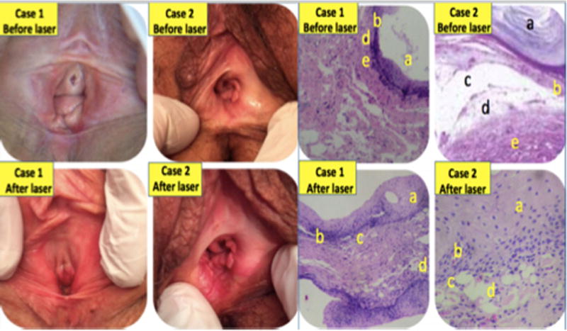
Vulvar lichen sclerosus. Histological assessment of Masson’s trichrome-stained tissue samples before versus after factional CO2 laser treatment. Left, case 1; Right, case 2. Histology: Top row: Hyperkeratosis (a), dermal atrophy, hydropic degeneration of the basal epithelial cells (b) dermo epidermal clefts (c) homogeneous papillary dermis, afibrillar with frosted glass appearance and edema (d) and inflammatory infiltrate of polymorphonuclear band and plasma cells (e) and inflammatory infiltrate of polymorphonuclear band and plasma cells. Bottom row: Trophic epithelium without superficial hyperkeratosis (a), persistence in some basal cells hydropic degeneration (b), persistence of some areas with dermo-epidermal clefts (c), and lamina propria fibrillar with irregular spaces containing translucent material (d). (Courtesy: Elias J, Galich M).
Vulvodynia and Provoked Vestibulodynia
Vulvodynia is defined as “pain in the vulva” persisting for at least 3 months, in the absence of an obvious underlying cause, and can include various clinical features. Some patients have continuous, diffuse vulvar pain (generalized, spontaneous vulvodynia), while others experience localized pain, usually provoked by touch. The current nomenclature [92] uses a symptom-based classification to characterize the pain in regard to its location (localized, generalized, mixed), conditions that provoke it (contact, spontaneous, or mixed), its temporal pattern (intermittent or constant), and its onset (primary or secondary).
Provoked, localized vestibulodynia (PVD), previously called “vulvar vestibulitis syndrome,” is the term used to describe the phenomena of superficial pain confined to the vulvar vestibule, provoked by touch. In some women, pain can be caused by minimal touch (sitting, tight-fitting clothing), whereas in others, it is provoked by vaginal penetration during sexual intercourse, tampon insertion, or gynecological examination, resulting in dyspareunia or complete inability to have intercourse. The pain is often described as burning or cutting and can radiate throughout the entire vestibule or confined to the lower vestibule. Erythema may be evident but is no longer considered a defining criterion.
No single causative factor has yet been identified, and the etiology is considered multifactorial [93]. Clinical observations, epidemiological studies, and basic research suggest different etiologies, including inflammation, hormonal factors, pelvic floor muscle dysfunction, peripheral and central neural pain disorders, genetic predisposition, psychosocial, and sexual factors. Current diagnosis is based upon clinical symptoms while treatment is assessed on a trial and error approach. As a result, many forms of therapeutic interventions have been used, and outcomes remain largely inconclusive, with response rate ranging between 40% and 85% and with many women not responding to any treatment [94]. Given the high placebo response rate of around 50%, only placebo-controlled trials will provide substantial data about beneficial therapeutic techniques. In a recent study patients (N= 70) underwent fractional micro-ablative CO2 laser treatment for vestibular pain plus vestibulodynia (n = 37) or GSM (N = 33). A statistically significant improvement of symptoms was noted after three sessions of vestibular fractional CO2 laser treatment. Improvements gradually increased throughout the study period and were maintained through the 4 month follow-up period [95].
External Genitals
Energy based devices are used to perform surgical procedures on the external genitals: some are for medical indications and some are considered as aesthetic gynecology. Treatment outcome on the use of low level laser therapy on episiotomy [Alvarenga A. et al.] and fractional CO2 laser on cesarean section scars [Karmisholt K. et al] are published in this issue. The field of aesthetic gynecology lies beyond the scope of this review and some highlights are briefly mentioned. (Table 3) [96–98].
TABLE 3.
Vaginal Rejuvenation for Enhancement of Sexual Gratification
| Author | Indication | N | Device | Findings | Complic. |
|---|---|---|---|---|---|
| Millheiser et al. (2010) [96] | Laxity after childbirth | 24 at 3 months | Monopolar RF | All subjects with improved vaginal tightness at 1 month; sustained or further improved by 3 months | None |
| Lee (2014) [97] | Laxity | 30 at 2 months | Er:YAG | Perineometer values improved in all patients, 70% subjective improvement | None |
| Vicariotto and Raichi (2016) [98] | Laxity | 11 at 2 months (premenopausal, vaginal laxity arm). 12 at 2 months (postmenopausal, VVA/GSM arm) | Dynamic quadripolar radiofrequency (DQRF) | Improvement reported at 4–8 weeks in laxity and urinary symptoms, VVA symptoms, and sexual satisfaction | None |
Hopes or Hypes
Randomized controlled trials (RCT), peer-reviewed publications, meta-analyses, and other objective evaluations all aim to protect patients from a medical procedure that has not been adequately tested and proven safe and effective. This review, drafted by a large group of experts may lack some of the above-mentioned “protective tools,” but we hope that it offers relevant collective information to define directions for future research, and may serve as “The Guide to the Perplexed” in the arena of gynecologic energy-based clinical procedures.
A gynecologist’s office serves as a refuge and, occasionally, as a confessional when discussing the most private gynecologic, sexual and other intimate concerns. Surgeons discerningly consider behavioral, medical, pharmacologic, and reconstructive pelvic surgical procedures, in addition to potential medical and cosmetic external treatments. Office-based cosmetic procedures offered by plastic, dermatologic, or gynecologic surgeons provide solutions for common vulvovaginal disorders; however, it is unclear whether alternatives are always fully presented to women seeking cosmetic services. Form and function are both equally important for pelvic floor health as well as vulvovaginal health. Patients may seek cosmetic gynecological treatments for more than just aesthetic reasons. Functionally, they may struggle with clothing discomfort or dyspareunia [99] in addition to struggles relating to body image or feelings of inadequacy during intimate moments. Patients may also have complaints of vaginal laxity, now defined by the IUGA/ICS as “excessive vaginal looseness,” [100] or have issues with air trapping or vaginal wind resulting in noises emanating from the vagina during activity [101].
While vaginal laxity refers to loose vaginal tissue, this broad and unspecified term is subjective when contrasted with well-quantified pelvic floor disorders, including pelvic organ prolapse (cystocele, uterine prolapse, rectocele, and vaginal vault prolapse), SUI and fecal incontinence. Such disorders typically result from loss of connective tissue support in concert with muscle and nerve damage, most commonly occurring after childbirth or related to other inciting factors such as chronic cough and straining associated with constipation. Conversely, vaginal laxity and enlarged labia majora or minora are often a matter of personal preference which can be influenced by the media, social media, and relationships [102–106]. While numerous case series have been published on labiaplasty/vaginoplasty outcomes [103–106], no comparative trials exist on labiaplasty techniques and their clinical efficacy in improving orgasmic response. Risks related to clitoral and labial surgery include denervation and scarring. Procedures such as re-virgination represent lack of education and misinformation perpetuating myths that virginity can be confirmed by the presence/absence of a hymen.
Data relating to energy-based therapies in cosmetogynecology primarily focus on treatment of vulvovaginal atrophy/GSM. Unfortunately, the large majority of the trials are retrospective, single-center studies with short-term follow-up. Furthermore, outcomes measures are not standardized and are often only subjective or histology-based. Energy-based devices are currently being employed for treatment of labial hypertrophy, vaginal laxity, and reduced sexual pleasure, but limited published data are available.
The vaginal laser versus estrogen cream therapy (VELVET) trial, comparing the effects of these two treatment options of vaginal dryness, is currently recruiting GSM patients at the Cleveland Clinic and five other sites across the United States. The Vaginal Erbium Laser Academy Study (VELAS) [107] will examine vaginal erbium laser therapy as a potentially non-invasive treatment for GSM and for SUI. This international study will include 1,500 postmenopausal women. The CURLS study, aiming to assess clobetasol versus fractional CO2 laser for treatment of vulvar lichen sclerosus, is currently recruiting patients at Medstar Health Washington Hospital Center in Washington, DC. A trial on the effect of fractional/pixel CO2 laser on GSM symptoms is in progress at the Cleveland Clinic, Florida, and monitoring of tissue changes with advanced optical techniques is in under review at the Beckman Laser Institute, University of California, Irvine. Other studies with CO2 laser, Er:YAG laser, and RF are in progress in Italy, Greece, Argentina, Slovenia, Israel, Spain, the UK, UAE, Japan, Korea, Indonesia, and more sites around the globe.
Consensus and Controversies
Data generated from the unprecedented wave of publications in peer-review journals on cell activation and vaginal wall rejuvenation following exposure to energy-based devices justify the quotation: “You can fool all the people some of the time, and some of the people all the time, but you cannot fool all the people all the time” (Abraham Lincoln).
A consensus has been reached among the authors of this manuscript on the clinical effectiveness of energy-based therapies on GSM-related symptoms. The currently available data on the effects of fractional laser and RF on the skin, and additional information reported in almost 20 peer-reviewed publications on GSM-related symptoms, unequivocally demonstrate the following vaginal changes: thickening of glycogen enriched postmenopausal epithelium, neovascularization, and neocollagenesis in the lamina propria, increased lactobacilli counts, reduced pH, vaginal wall tightening, and improved urination control with minimal risk of short- and long-term complications.
Controversial issues that remain to be determined: the preferred energy-based device, be it laser versus RF, CO2 versus Er:YAG laser, tissue scanning versus pixel stamping of the fractional laser technology, ablative versus non-ablative technology, single laser wavelength versus hybrid consecutive beams, the optimal depth of micro-ablation or temperature of controlled energy deposition. In addition, understanding the physiologic cascade of events during the process of tissue rejuvenation, most effective treatment session intervals and duration of effects that justify a touch-up session remain to be determined. Studies are necessary to evaluate the possible curative effect on lichen sclerosus, vestibulitis, additive effect of platelet-rich plasma/growth factor, and hyaluronic acid when administered in conjunction with energy deposition. The potential impact of the energy-based treatment on estrogen receptors levels in the vaginal wall will also need to be addressed. Add-on therapies such as estrogen priming for vaginal atrophy (Gaspar A, et al. described elsewhere in this issue) [67], corticosteroids for lichen sclerosus, or superficial tissue cooling [108] for all of the above also need further investigation. Computer simulations of thermal tissue remodeling during transvaginal and transurethral laser treatment of female SUI [Hardy L. et al.] described elsewhere in this issue, and the potential use of intra-urethral fractional laser treatment of female SUI (Gaspar A. et al. unpublished data) are tested. The cost-effectiveness of energy-based regimens in comparison to other available treatments, and unwanted long-term effects of such interventions all remain to be defined.
Acknowledgments
The authors gratefully acknowledge the information received from energy-based device manufacturers as presented in Table 1, and Figure 7. No commercial source supported the collaboration and preparation of this manuscript.
Footnotes
Conflict of Interest Disclosures: All authors have completed and submitted the ICMJE Form for Disclosure of Potential Conflicts of Interest and have disclosed the following: Yona Tadir is a scientific consultant for Alma Lasers. Red Alinsod is a consultant for Thermi, receive royalty for ThermiVa, Cooper Surgical, Lone Star Retractor, and Coloplast. Consultant, Caldera Medical: Stock owner, Monarch Medical Products: Consultant, Royalty for Alinsod Surgical Equipment. Selih-Martinec K. is an invited speaker by Alma Lasers. Alberto Calligaro, Stefano Salvatore, and Nicola Zerbinati are scientific consultants for DEKA laser. All other co-authors: nothing to disclose. No commercial source supported the collaboration and preparation of this manuscript.
[Correction added on 24 February 2017, after first online publication: The spelling of Jorge E. Gaviria’s name and affiliation number were corrected.]
References
- 1.Kaplan I, Goldman J, Ger R. The treatment of erosions of the uterine cervix by means of the CO2 laser. Obstet Gynecol. 1973;41(5):795–796. [PubMed] [Google Scholar]
- 2.Bellina JH, Polanyi TG. Management of vaginal adenosis and related cervico-vaginal disorders in DES-exposed pregnancy by means of carbon dioxide laser surgery. J Reprod Med. 1976;16:295–296. [PubMed] [Google Scholar]
- 3.Bruhat M, Mage G, Mahnes M. Use of the CO2 laser in laparoscopy. Proceedings of the 3rd international society for laser surgery. Ed. Kaplan I. 1979;24:235–238. [Google Scholar]
- 4.Tadir Y, Kaplan I, Zukerman Z, Ovadia J. Laparoscopic CO2 laser sterilization. In: Semm K, Mettler L, editors. Human Reproduction. Exerpta Medica, Amsterdam: Elsevier; 1981. pp. 551pp. 423–431. [Google Scholar]
- 5.Nezhat FR, Nezhat CR, Silfen SL. Videolaseroscopy for oophorectomy. Am J Obstet Gynecol. 1991;165:1323–1330. doi: 10.1016/0002-9378(91)90360-4. [DOI] [PubMed] [Google Scholar]
- 6.Baggish MS, ElBakry MM. A flexible CO2 laser fiber for operative laparoscopy. Fertil Steril. 1986;46(1):16–20. doi: 10.1016/s0015-0282(16)49450-1. [DOI] [PubMed] [Google Scholar]
- 7.Cossmann PH, Romano V, Spörri S, et al. Plastic hollow waveguides: Properties and possibilities as a flexible radiation delivery system for CO2 laser radiation. Lasers Surg Med. 1995;16(1):66–75. doi: 10.1002/lsm.1900160109. [DOI] [PubMed] [Google Scholar]
- 8.Tadir Y, Kaplan I, Zukerman Z, Ovadia J. Hysteroscope for CO2 laser application. Lasers Surg Med. 1984;4:153–156. doi: 10.1002/lsm.1900040205. [DOI] [PubMed] [Google Scholar]
- 9.Haimovitch S, Lopez-Yarto M, Urresta-Avila J, Saaverda Tascon A, Hernandez JL, Carreras Collado R. Office hysteroscopic laser enucleation of sumcucous myomas without mass expraction: A case study. Biomed Res Int. 2015;2015:905204. doi: 10.1155/2015/905204. [DOI] [PMC free article] [PubMed] [Google Scholar]
- 10.Tadir Y, Wright WH, Vafa O, Ord T, Asch R, Berns MW. Micromanipulation of sperm with a laser generated optical trap. Fertil Steril. 1989;52:870–875. [PubMed] [Google Scholar]
- 11.Harper JC, Wilton L, Traeger-Synodinos J, et al. The ESHRE PGD consortium: 10 years of data collection. Hum Reprod Update. 2012;18(3):234–247. doi: 10.1093/humupd/dmr052. [DOI] [PubMed] [Google Scholar]
- 12.Brightman LA, Brauer JA, Anolik R, Weiss E, Karen J, Chapas A, Hale E, Bernstein L, Geronemus RG. Ablative and fractional ablative lasers. Dermatol Clin. 2009;27(4):479–489. doi: 10.1016/j.det.2009.08.009. [DOI] [PubMed] [Google Scholar]
- 13.Gaspar A, Addamo G, Brandi H. Vaginal fractional CO2 laser: A minimally invasive option for vaginal rejuvenation. Am J Cosmetic Surg. 2011;28(3):156–162. [Google Scholar]
- 14.Salvatore S, Leone Roberti Maggiore U, Athanasiou S, et al. Histological study on the effect of microablative fractional CO2 laser on atrophic vaginal tissue: An ex vivo study. Menopause. 2015;22(8):845–849. doi: 10.1097/GME.0000000000000401. [DOI] [PubMed] [Google Scholar]
- 15.Zerbinati N, Serati M, Origoni M, et al. Microscopic and ultrastructural modifications of postmenopausal atrophis vaginal mucosa after fractional carbon dioxide laser treatment. Lasers Med Sci. 2015;30(1):429–436. doi: 10.1007/s10103-014-1677-2. [DOI] [PubMed] [Google Scholar]
- 16.Reid R, Absten GT. Lasers in gynecology: Why pragmatic surgeons have not abandoned this valuable technology. Lasers Surg Med. 1995;17(4):378–383. doi: 10.1002/lsm.1900170302. [DOI] [PubMed] [Google Scholar]
- 17.Copperman AB, DeCherney AH. Turn, turn, turn. Fertil Steril. 2006;85(1):12–13. doi: 10.1016/j.fertnstert.2005.07.1297. [DOI] [PubMed] [Google Scholar]
- 18.De Landsheere L, Munaut C, Nusgens B, et al. Histology of the vaginal wall in women with pelvic organ prolapse: A literature review. Int Urogynecol J. 2013;24(12):2011–2020. doi: 10.1007/s00192-013-2111-1. [DOI] [PubMed] [Google Scholar]
- 19.Jannini EA, d’Amati G, Lenzi A. Histology and immunohistochemical studies of female genital tissue. In: Goldstein I, Meston CM, Davis S, Traish A, editors. Women’s Sexual Function and Dysfunction: Study, Diagnosis and Treatment. UK: CBC Press, Taylor an Francis Group; 2005. p. 784. [Google Scholar]
- 20.Wenstrup RJ, Florer JB, Brunskill EW, Bell SM, Chervoneva I, Birk DE. Type V collagen controls the initiation of collagen fibril assembly. J Biol Chem. 2004;279(51):53331–53337. doi: 10.1074/jbc.M409622200. [DOI] [PubMed] [Google Scholar]
- 21.Birk DE, Fitch JM, Babiarz JP, Doane KJ, Linsenmayer TF. Collagen fibrillogenesis in vitro: Interaction of types I and V collagen regulates fibril diameter. J Cell Sci. 1990;95(4):649–657. doi: 10.1242/jcs.95.4.649. [DOI] [PubMed] [Google Scholar]
- 22.Menopause society. Management of symptomatic vulvovaginal atrophy: 2013 position statement of The North American Menopause Society. Menopause. 2013;20(9):888–902. 904. doi: 10.1097/GME.0b013e3182a122c2. [DOI] [PubMed] [Google Scholar]
- 23.Cotreau MM, Chennathukuzhi VM, Harris HA, et al. A study of 17beta-estradiol-regulated genes in the vagina of postmenopausal women with vaginal atrophy. Maturitas. 2007;58(4):366–376. doi: 10.1016/j.maturitas.2007.09.009. [DOI] [PubMed] [Google Scholar]
- 24.Portman DJ, Gass MLS. Genitourinary syndrome of menopause: New terminology for vulvovaginal atrophy from the International Society for the Study of Women’s Sexual Health and The North American Menopause Society. Maturitas. 2014;79(3):349–354. doi: 10.1016/j.maturitas.2014.07.013. and Menopause 2014;21 (10):1063–1068. [DOI] [PubMed] [Google Scholar]
- 25.Willhite LA, O’Connell MB. Urogenital atrophy: Prevention and treatment. Pharmacotherapy. 2001;21(4):464–480. doi: 10.1592/phco.21.5.464.34486. [DOI] [PubMed] [Google Scholar]
- 26.Bachmann G. Clinical manifestations and diagnosis of vaginal atrophy. UpToDate. 2014:1–10. [Google Scholar]
- 27.Alexiades MR. Wave science in dermatologic therapy. J Drugs Dermatol. 2015;14(11):1190. [PubMed] [Google Scholar]
- 28.Alexiades-Armenakas MR, Dover JS, Arndt KA. The spectrum of laser skin resurfacing: Nonablative, fractional, and ablative laser resurfacing. J Am Acad Dermatol. 2008;58–(5):719–737. 738–40. doi: 10.1016/j.jaad.2008.01.003. [DOI] [PubMed] [Google Scholar]
- 29.Fisher JC. Photons, physiatrics, and physicians: A practical guide to understanding laser light interaction with living tissue, part 1. J Clin Laser Med Surg. 1992;10(6):419–426. doi: 10.1089/clm.1992.10.419. [DOI] [PubMed] [Google Scholar]
- 30.Anderson R, Parrish JA. Selective photothermolysis: Precise microsurgery by selective absorption of pulsed radiation. Science, New Series. 1983;220(4596):524–527. doi: 10.1126/science.6836297. [DOI] [PubMed] [Google Scholar]
- 31.Alexiades-Armenakas MR, Dover JS, Arndt KA. Fractional laser skin resurfacing. J Drugs Dermatol. 2012;11(11):1274–1287. [PubMed] [Google Scholar]
- 32.Alexiades-Armenakas M, Sarnoff D, Gotkin R, Sadick N. Multi-center clinical study and review of fractional ablative CO2 laser resurfacing for the treatment of rhytides, photoaging, scars and striae. J Drugs Dermatol. 2011;10(4):352–362. [PubMed] [Google Scholar]
- 33.Baker-Jarvis J, Kim S. The interaction of radio-frequency fields with dielectric materials at macroscopic to mesoscopic scales. J Res Inst Stand Technol. 2012;117:1–60. doi: 10.6028/jres.117.001. [DOI] [PMC free article] [PubMed] [Google Scholar]
- 34.Alexiades M, Berube D. Randomized, blinded, 3-arm clinical trial assessing optimal temperature and duration for treatment with minimally invasive fractional radiofrequency. Dermatol Surg. 2015;41(5):623–632. doi: 10.1097/DSS.0000000000000347. [DOI] [PubMed] [Google Scholar]
- 35.Dunbar SW, Goldberg DJ. Cosmetic dermatology: An update. J Drug Dermatol. 2015;14(11):1229–1238. [PubMed] [Google Scholar]
- 36.Alexiades-Armenakas M, Dover JS, Arndt KA. Unipolar versus bipolar radiofrequency treatment of rhytides and laxity using a mobile painless delivery method. Lasers Surg Med. 2008;40(7):446–453. doi: 10.1002/lsm.20667. [DOI] [PubMed] [Google Scholar]
- 37.Lee MS. Treatment of vaginal relaxation syndrome with an Erbium:YAG laser using 90° and 360° scanning scopes: A pilot study & short-term results. Laser Ther. 2014;23(2):129–138. doi: 10.5978/islsm.14-OR-11. [DOI] [PMC free article] [PubMed] [Google Scholar]
- 38.Ogrinc UB, Sencar S, Lenasi H. Novel minimally invasive laser treatment of urinary incontinence in women. Lasers Surg Med. 2015;47:689–697. doi: 10.1002/lsm.22416. [DOI] [PMC free article] [PubMed] [Google Scholar]
- 39.Fistonic N, Fistonic I, Gustek SF, et al. Minimally invasive, non-ablative Er:YAG laser treatment of stress urinary incontinence in women: A pilot study. Laser Med Sci. 2016;31:635–643. doi: 10.1007/s10103-016-1884-0. [DOI] [PMC free article] [PubMed] [Google Scholar]
- 40.Alexiades-Armenakas M, Newman J, Willey A, Kilmer S, et al. Prospective multicenter clinical trial of a minimally-invasive temperature-controlled bipolar fractional radiofrequency system for rhytid and laxity treatment. Dermatol Surg. 2013;39(2):263–273. doi: 10.1111/dsu.12065. [DOI] [PubMed] [Google Scholar]
- 41.Saedi N, Jalian HR, Petelin A, Zachary C. Fractionation: Past, present, future. Semin Cutan Med Surg. 2012;31(2):105–109. doi: 10.1016/j.sder.2012.02.003. [DOI] [PubMed] [Google Scholar]
- 42.Capon A, Mordon A. Can thermal laser promote skin wound healing? Am J Clin Dermatol. 2003;4(1):1–12. doi: 10.2165/00128071-200304010-00001. [DOI] [PubMed] [Google Scholar]
- 43.Lev-Sagie A. Vulvar and vaginal atrophy: Physiology, clinical presentation, and treatment considerations. Clin Obstet Gynecol. 2015;58(3):476–491. doi: 10.1097/GRF.0000000000000126. [DOI] [PubMed] [Google Scholar]
- 44.Berlin AL, Hussain M, Phelps R, Goldberg DJ. A prospective study of fractional scanned nonsequential carbon dioxide laser resurfacing: A clinical and histopathologic evaluation. Dermatol Surg. 2009;35(2):222–228. doi: 10.1111/j.1524-4725.2008.34413.x. [DOI] [PubMed] [Google Scholar]
- 45.Lee CJ, Park JH, Ciesielski TE, Thomson JG, Persing JA. Retinoids, 585-nm laser, and carbon dioxide laser: A numeric comparison of neocollagen formation in photoaged hairless mouse skin. Aesth Plast Surg. 2008;32(6):894–901. doi: 10.1007/s00266-008-9121-2. [DOI] [PubMed] [Google Scholar]
- 46.Salvatore S, Nappi RE, Zerbinati N, et al. A 12-week treatment with fractional CO2 laser for vulvovaginal atrophy: A pilot study. Climacteric. 2014;17(4):363–369. doi: 10.3109/13697137.2014.899347. [DOI] [PubMed] [Google Scholar]
- 47.Perino A, Calligaro A, Forlani F, et al. Vulvo-vaginal atrophy: A new treatment modality using thermoablative fractional CO2 laser. Maturitas. 2015;80:296–301. doi: 10.1016/j.maturitas.2014.12.006. [DOI] [PubMed] [Google Scholar]
- 48.Sokol ER, Karram M. An assessment of the safety and efficacy of a fractional CO2 laser system for the treatment of vulvovaginal atrophy. Menopause. 2016;23(10):1102–1107. doi: 10.1097/GME.0000000000000700. [DOI] [PubMed] [Google Scholar]
- 49.Leone Roberti Maggiore U, Parma M, Candiani M, Salvatore S. Microablative fractional CO2 laser for vulvo vaginal atrophy in women with an history of breast cancer. J Minim Invasive Gynecol. 2015;22(6S):S100. doi: 10.1016/j.jmig.2015.08.269. [DOI] [PubMed] [Google Scholar]
- 50.Pieralli A, Fallani MG, Becorpi A, et al. Fractional CO2 laser for vulvovaginal atrophy (VVA) dyspareunia relief in breast cancer survivors. Arch Gynecol Obstet. 2016;294(4):841–846. doi: 10.1007/s00404-016-4118-6. [DOI] [PubMed] [Google Scholar]
- 51.Pagano T, De Rosa P, Vallone R, et al. Fractional micro-ablative CO2 laser for vulvovaginal atrophy in women treated with chemotherapy and/or hormonal therapy for breast cancer: A retrospective study. Menopause. 2016;23(10):1108–1113. doi: 10.1097/GME.0000000000000672. [DOI] [PubMed] [Google Scholar]
- 52.Mac Bride MB, Rhodes DJ, Shuster LT. Vulvovaginal atrophy. Mayo Clin Proc. 2010;85(1):87–94. doi: 10.4065/mcp.2009.0413. [DOI] [PMC free article] [PubMed] [Google Scholar]
- 53.Athanasiou S, Pitsouni E, Antonopoulou S, et al. The effect of microablative fractional CO2 laser on vaginal flora of postmenopausal women. Climacteric. 2016;19(5):512–518. doi: 10.1080/13697137.2016.1212006. [DOI] [PubMed] [Google Scholar]
- 54.Stephens DJ. Cell biology: Collagen secretion explained. Nature. 2012;482(7386):474–475. doi: 10.1038/482474a. [DOI] [PMC free article] [PubMed] [Google Scholar]
- 55.Alberts B, Johnson A, Lewis J. Transport from the ER through the Golgi apparatus Molecular Biology of the Bell. 4th edition. New York: Garland Science; 2002. pp. 775–781. [Google Scholar]
- 56.Orringer JS, Kang S, Johnson TJ, et al. Connective tissue remodeling induced by carbon dioxide laser resurfacing of photodamaged human skin. Arch Dermatol. 2004;140(11):1326–1332. doi: 10.1001/archderm.140.11.1326. [DOI] [PubMed] [Google Scholar]
- 57.Hantash BM, Bedi VP, Kapadia B, et al. In vivo histological evaluation of a novel ablative fractional resurfacing device. Lasers Surg Med. 2007;39:96–107. doi: 10.1002/lsm.20468. [DOI] [PubMed] [Google Scholar]
- 58.Ishida Y, Nagata K. Hsp47 as a collagen-specific molecular chaperone. Methods Enzymol. 2011;499:167–182. doi: 10.1016/B978-0-12-386471-0.00009-2. [DOI] [PubMed] [Google Scholar]
- 59.Tasab M, Batten MR, Bulleid NJ. Hsp47: A molecular chaperone that interacts with and stabilizes correctly-folded procollagen. EMBO J. 2000;19(10):2204–2211. doi: 10.1093/emboj/19.10.2204. [DOI] [PMC free article] [PubMed] [Google Scholar]
- 60.Dafforn TR, Della M, Miller AD. The molecular interactions of heat shock protein 47 (Hsp47) and their implications for collagen biosynthesis. J Biol Chem. 2001;276(52):49310–49319. doi: 10.1074/jbc.M108896200. [DOI] [PubMed] [Google Scholar]
- 61.Prignano F, Campolmi P, Bonan P, et al. Fractional CO2 laser: A novel therapeutic device upon photobiomodulation of tissue remodeling and cytokine pathway of tissue repair. Dermatol Ther. 2009;22(Suppl 1):S8–S15. doi: 10.1111/j.1529-8019.2009.01265.x. [DOI] [PubMed] [Google Scholar]
- 62.Nowak KC, McCormack M, Koch RJ. The effect of superpulsed carbon dioxide laser energy on keloid and normal dermal fibroblast secretion of growth factors: A serum-free study. Plast Reconstr Surg. 2000;105(6):2039–2048. doi: 10.1097/00006534-200005000-00019. [DOI] [PubMed] [Google Scholar]
- 63.Manolis EN, Kaklamanos IG, Spanakis N, et al. Tissue concentration of transforming growth factor b1 and basic fibroblast growth factor in skin wounds created with a CO2 laser and scalpel: A comparative experimental study, using an animal model of skin resurfacing. Wound Repair Regen. 2007;15:252–257. doi: 10.1111/j.1524-475X.2007.00212.x. [DOI] [PubMed] [Google Scholar]
- 64.Reinisch L. Scatter-limited phototherapy: A model for laser treatment, of skin. Lasers Surg Med. 2002;30(5):381–388. doi: 10.1002/lsm.10046. [DOI] [PubMed] [Google Scholar]
- 65.Lukaö M, Mqjaron B, Rupnik T. Ablative and thermal effects of Er:YAG laser on human tissue. In: Waidelich W, Waidelich R, Waldschmidt J, editors. Lasers in Medicine. Berlin: Springer; 1998. pp. 566–572. [Google Scholar]
- 66.Majaron B, Plesteniak Lukaé M. Thermo-mechanical laser ablation of soft biological tissue: Modeling the micro explosions. Appl Phys B. 1999;69:71–80. [Google Scholar]
- 67.Gaspar A, Brandi H, Gomez V, Luque D. Efficacy of Erbium: YAG laser treatment compared to topical estriol treatment for symptoms of genitourinary syndrome of menopause. Lasers Surg Med. 2016 doi: 10.1002/lsm.22569. [DOI] [PMC free article] [PubMed] [Google Scholar]
- 68.Masters WH, Johnson VE. Human Sexual Response. Toronto; New York: Bantam; Brooks: 1966. [Google Scholar]
- 69.Millheiser LS, Pauls RN, Herbst SJ, Chen BH. Radio-frequency treatment of vaginal laxity after vaginal delivery: Nonsurgical vaginal tightening. J Sex Med. 2010;7(9):3088–3095. doi: 10.1111/j.1743-6109.2010.01910.x. [DOI] [PubMed] [Google Scholar]
- 70.Mulholland RS. Radio frequency energy for non-invasive and minimally invasive skin tightening. Clin Plast Surg. 2011;38(3):437–448. doi: 10.1016/j.cps.2011.05.003. [DOI] [PubMed] [Google Scholar]
- 71.Alinsod RM. Transcutaneous temperature controlled radio-frequency for orgasmic dysfunction. Lasers Surg Med. 2016;48(7):641–645. doi: 10.1002/lsm.22537. [DOI] [PMC free article] [PubMed] [Google Scholar]
- 72.Metelitsa AI, Alster TS. Fractionated laser skin resurfacing treatment complications: A review. Dermatol Surg. 2010;36(3):299–306. doi: 10.1111/j.1524-4725.2009.01434.x. [DOI] [PubMed] [Google Scholar]
- 73.Norton P, Brubaker L. Urinary incontinence in women. Lancet. 2006;367:57–67. doi: 10.1016/S0140-6736(06)67925-7. [DOI] [PubMed] [Google Scholar]
- 74.Fistonic N, Fistonic I, Lukanovic A, Findri-Gustek S, Sorta Bilajac Turina I, Franic D. First assessment of short term efficacy of Er:YAG laser treatment on stress urinary incontinence in women: Prospective cohort study. Climacteric. 2015;18(Sup 1):37–42. doi: 10.3109/13697137.2015.1071126. [DOI] [PubMed] [Google Scholar]
- 75.Ogrinc BU, Sencar S. Novel minimally invasive laser treatment of urinary incontinence in women. Lasers Surg Med. 2015;47:689–697. doi: 10.1002/lsm.22416. [DOI] [PMC free article] [PubMed] [Google Scholar]
- 76.Fistonic N, Fistonic I, Findri Gustek SS, et al. Minimally invasive, non-ablative Er:YAG laser treatment of stress urinary incontinence in women—A pilot study. J Laser Med Sci. 2016;31:635–643. doi: 10.1007/s10103-016-1884-0. [DOI] [PMC free article] [PubMed] [Google Scholar]
- 77.Pardo J, Sola V, Morales A. Treatment of female stress urinary incontinence with Erbium YAG laser in non-ablative mode. Eur J Obstet Gynecol Reprod Biol. 2016;204:1–4. doi: 10.1016/j.ejogrb.2016.06.031. [DOI] [PubMed] [Google Scholar]
- 78.Tien YW, Hsiao SM, Lee CN, Lin HH. Effects of laser procedure for female urodynamic stress incontinence on pad weight, urodynamics, and sexual function. Int Urogynecol J. 2016 doi: 10.1007/s00192-016-3129-y. Epub ahead of print. [DOI] [PubMed] [Google Scholar]
- 79.Pitsouni E, Grigoriadis T, Tsiveleka A, et al. Microablative fractional CO2-laser therapy and the genitourinary syndrome of menopause: An observational study. Maturitas. 2016;94:131–136. doi: 10.1016/j.maturitas.2016.09.012. [DOI] [PubMed] [Google Scholar]
- 80.Leibaschoff G, Izasa PG, Cardona JL, Miklos JR, Moore RD. Transcutaneous temperature controlled radiofrequency (TTCRF) for the treatment of menopausal vaginal/genitourinary symptoms. Surg Technol Int. 2016;26(XXIX):149–159. [PubMed] [Google Scholar]
- 81.Mirowski GW, Edwards L. Geniatl anatomy. In: Edwards L, editor. Genital Dermatology Atlas. Philadelphia: Lippincott Williams and Wilkins; 2004. pp. 1–8. [Google Scholar]
- 82.Habif TP. Skin anatomy. In: Mosby, editor. Clinical Dermatology. St. Louis, MI: 2004. [Google Scholar]
- 83.Smith YR, Haefner HK. Vulvar lichen sclerosus: Pathophysiology and treatment. Am J Clin Dermatol. 2004;5(2):105–125. doi: 10.2165/00128071-200405020-00005. [DOI] [PubMed] [Google Scholar]
- 84.Goldstein AT, Marinoff SC, Christopher K, Srodon M. Prevalence of vulvar lichen sclerosus in a general gynecology practice. J Reprod Med. 2005;50(7):477. [PubMed] [Google Scholar]
- 85.Cooper SM, Arnold SJ. Vulvar lichen sclerosus. uptodate 2016, last updated: Jun 06, 2016. Available at: http://www.uptodate.com/
- 86.Bradford J, Fischer G. Long-term management of adult vulvar lichen sclerosus: A prospective cohort study of 507 women. JAMA Dermatol. 2015;151(10):1061–1067. doi: 10.1001/jamadermatol.2015.0643. [DOI] [PubMed] [Google Scholar]
- 87.Kartamaa M, Reitamo S. Treatment of lichen sclerosus with carbon dioxide laser vaporization. Br J Dermatol. 1997;136(3):356–359. [PubMed] [Google Scholar]
- 88.Windahl T. Is carbon dioxide laser treatment of lichen sclerosus effective in the long run? Scand J Urol Nephrol. 2006;40(3):208–211. doi: 10.1080/00365590600589666. [DOI] [PubMed] [Google Scholar]
- 89.Peterson C, Lane J, Ratz J. Successful carbon dioxide laser therapy for refractory anogenital lichen sclerosus. Dermatol Surg. 2004;30:1148–1151. doi: 10.1111/j.1524-4725.2004.30343.x. [DOI] [PubMed] [Google Scholar]
- 90.Lee A, Lim A, Fischer G. Fractional carbon dioxide laser in recalcitrant vulval lichen sclerosus. Australas J Dermatol. 2016;57(1):39–43. doi: 10.1111/ajd.12305. [DOI] [PubMed] [Google Scholar]
- 91.Baggish M. Fractional CO2 laser treatment for vaginal atrophy and vulvar lichen sclerosus. J Gynecol Surg. 2016;32(6):309–317. [Google Scholar]
- 92.Bornstein J, Goldstein AT, Stockdale CK, et al. ISSVD, ISSWSH and IPPS consensus terminology and classification of persistent vulvar pain and vulvodynia. Obstet Gynecol. 2016;127(4):745–751. doi: 10.1097/AOG.0000000000001359. [DOI] [PubMed] [Google Scholar]
- 93.Pukall CF, Goldstein AT, Bergeron S, et al. Vulvodynia: Definition, prevalence, impact, and pathophysiological factors. J Sex Med. 2016;13(3):291–304. doi: 10.1016/j.jsxm.2015.12.021. [DOI] [PubMed] [Google Scholar]
- 94.Andrews JC. Vulvodynia interventions—Systemic review and evidence grading. Obstet Gynecol Surv. 2011;66(5):299–315. doi: 10.1097/OGX.0b013e3182277fb7. [DOI] [PubMed] [Google Scholar]
- 95.Murina F, Karram M, Salvatore S, Felice R. Fractional CO2 laser treatment of the vestibule for patients with vestibulodynia and genitourinary syndrome of menopause: A pilot study. J Sex Med. 2016;13(12):1915–1917. doi: 10.1016/j.jsxm.2016.10.006. [DOI] [PubMed] [Google Scholar]
- 96.Millheiser LS, Pauls RN, Herbst SJ, Chen BH. Radio-frequency treatment of vaginal laxity after vaginal delivery: Nonsurgical vaginal tightening. J Sex Med. 2010;7(9):3088–3095. doi: 10.1111/j.1743-6109.2010.01910.x. [DOI] [PubMed] [Google Scholar]
- 97.Lee MS. Treatment of vaginal relaxation syndrome with an Erbium:YAG laser using 90° and 360° scanning scopes: A pilot study & short-term results. Laser Ther. 2014;23(2):129–138. doi: 10.5978/islsm.14-OR-11. [DOI] [PMC free article] [PubMed] [Google Scholar]
- 98.Vicariotto F, Raichi M. Technological evolution in the radio-frequency treatment of vaginal laxity and menopausal vulvo-vaginal atrophy and other genitourinary symptoms: First experiences with a novel dynamic quadripolar device. Minerva Ginecol. 2016;68(3):225–236. [PubMed] [Google Scholar]
- 99.Goodman MP, Otto PJ, Matlock DL, et al. Evaluation of body image and sexual satisfaction in women undergoing female genital plastic/cosmetic surgery. Aesthet Surg J. 2016;36(9):1048–1057. doi: 10.1093/asj/sjw061. [DOI] [PubMed] [Google Scholar]
- 100.Haylen BT, Maher CF, Barber MD, et al. An International Urogynecological Association (IUGA)/International Continence Society (ICS) joint report on the terminology for female pelvic organ prolapse (POP) Int Urogynecol J. 2016;27(2):165–194. doi: 10.1007/s00192-015-2932-1. [DOI] [PubMed] [Google Scholar]
- 101.Miranne J, Marek TM, Mete M, Iglesia CB. Prevalence and resolution of auditory passage of vaginal air in women with pelvic floor disorders. Obstet Gynecol. 2015;126(1):136–143. doi: 10.1097/AOG.0000000000000921. [DOI] [PubMed] [Google Scholar]
- 102.Cartwright R, Cardozo L. FOR: Women should be free to opt for cosmetic genital surgery. BJOG. 2014;121(6):767. doi: 10.1111/1471-0528.12622. [DOI] [PubMed] [Google Scholar]
- 103.Iglesia CB. AGAINST: The social vulnerability and cultural view of women as sex objects need to end. BJOG. 2014;121(6):768. doi: 10.1111/1471-0528.12621. [DOI] [PubMed] [Google Scholar]
- 104.Creighton S. AGAINST: Labiaplasty is an unnecessary cosmetic procedure. BJOG. 2014;121(6):768. doi: 10.1111/1471-0528.12620. [DOI] [PubMed] [Google Scholar]
- 105.Iglesia CB, Yurteri-Kaplan L, Alinsod R. Female genital cosmetic surgery: A review of techniques and outcomes. Int Urogynecol J. 2013;24(12):1997–2009. doi: 10.1007/s00192-013-2117-8. [DOI] [PubMed] [Google Scholar]
- 106.Sharp G, Tiggemann M, Mattiske J. Predictors of consideration of labiaplasty: An extension of the tripartite influence model of beauty ideals. Psychol Women Q. 2014:182–193. [Google Scholar]
- 107.Gambacciani M, Torelli MG, Martella L, et al. Rationale and design for the Vaginal Erbium Laser Academy Study (VELAS): An international multicenter observational study on genitourinary syndrome of menopause and stress urinary incontinence. Climacteric. 2015;18(Sup1):43–48. doi: 10.3109/13697137.2015.1071608. [DOI] [PubMed] [Google Scholar]
- 108.Nelson JS, Milner TE, Anvari B, Tenenbaum BS, Svaasand LO, Kimel S. Dynamic epidermal cooling in conjunction with laser-induced photothermolysis of port wine stain blood vessels. Lasers Surg Med. 1996;19(2):224–229. doi: 10.1002/(SICI)1096-9101(1996)19:2<224::AID-LSM15>3.0.CO;2-6. [DOI] [PubMed] [Google Scholar]