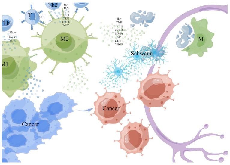
Figure 2.
Monocytes/macrophages. The M1 cell was activated by Th1 cytokine interferon-γ (IFN-γ), interleukin-12 (IL12), tumor necrosis factor (TNF), and microbial products, while the M2 cell was activated and differentiated by Th2 cytokines (such as IL4, IL5, IL10, IL13, colony-stimulating factor-1 (CSF1), transforming growth factor-1 (TFGβ1) and prostaglandin E2 (PGE2). TAMs (generally considered to be M2) exposed to hypoxia or lactate secretes a variety of cytokines with metabolic functions, including IL6, TNF, CCL5, and CCL18 to enhance PNI. TAMs express PD-L1 and can directly reduce the activation of T cells, then suppress the anticancer immune responses. TAMs promote tumor progression and invasion by up-regulating matrix metalloproteinases (MMPs). TAMs degrade the protein of nerve bundle membrane by expressing cathepsin B-mediated process. This picture is drawn by Figdraw (www.figdraw.com).

