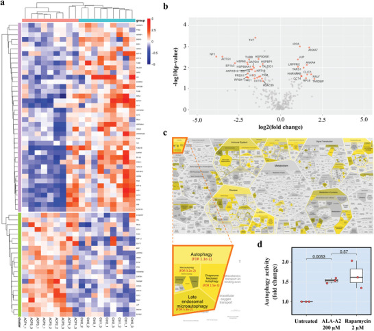
Figure 5.

SWATH‐proteomic and functional analyses suggest that cell death is caused by autophagic induction. a) Heatmap and b) volcano plot identified the significantly differentially expressed proteins between ALA‐A2 peptide‐treated versus untreated A549 cells (n = 9 per group; three technical replicates of three biological specimens) (full expression datasets in Table S2, Supporting Information). c) This reactome was subjected to biological pathway analyses. The stronger yellow accent indicates lower false‐discovery rate. Chaperone mediated autophagy was predicted as the most significant pathway elicited in ALA‐A2 peptide‐treated A549 cells (details in Table S3, Supporting Information). d) Autophagy activity assay. Rapamycin, an autophagic inducer, was the positive control. This experiment was performed in three biological replicates. *p < 0.05.
