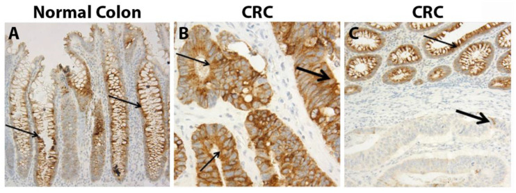
Figure 2.
IHC of MUC4 from Shanmugam et al., Cancer 2010, reprinted with permission [78]. (A) MUC4 staining was noted in crypts of the normal colon. (B) Strong cytoplasmic staining within CRC. (C) Weak staining of CRC compared to adjacent normal tissue. Thin arrows—normal colonic epithelium; thick arrows—CRC. Scale: (A) 200 μm; (B) 600 μm; (C) 200 μm.

