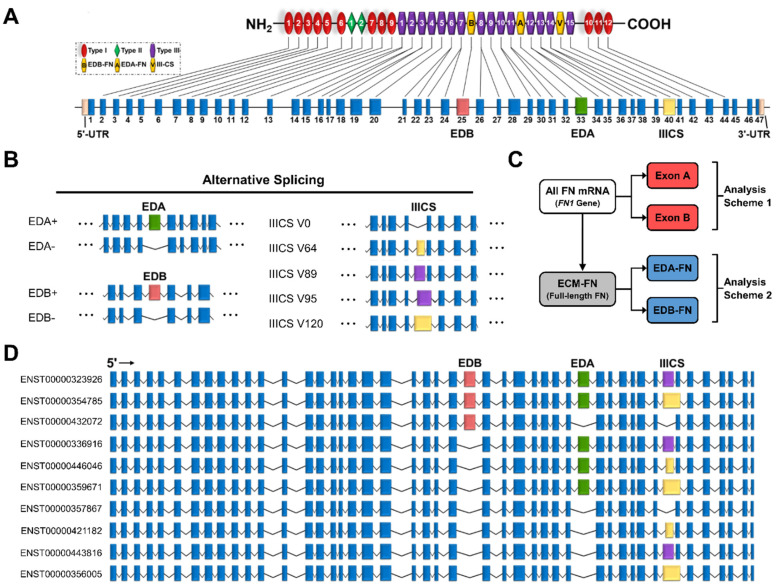
Figure 2.
Transcriptome of the FN1 gene. (A) FN protein and exon structure. The untranslated regions and exons that exhibit alternative splicing are in colors other than blue. (B) Alternative splicing of the EDA, EDB, and IIICS exons. The IIICS exon exhibits five splice variants of differing amino acid length indicated in the splice variant names. (C) Scheme displaying how transcripts are grouped for analysis. Exon A and Exon B are compared directly with all FN mRNAs (FN1 genes), while EDA-FNs and EDB-FNs are compared directly with ECM-FNs. (D) Exon structures of the 10 full-length ECM-FNs recognized in the Ensembl library.

