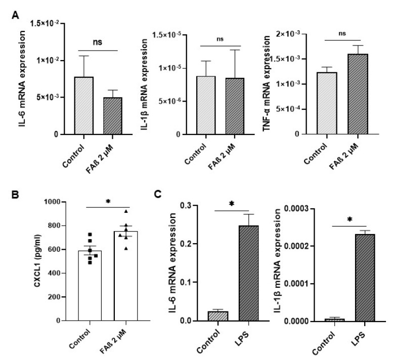
Figure 9.
Expression of inflammatory markers in astrocyte cells treated with FAβ1–42 peptides solution. (A) Three days post-stimulation with FAβ solution (2 µM), the expression of IL-6, IL-1β, and TNF-α were quantified by RT-qPCR. (B) CXCL1 was quantified by ELISA. (C) Astrocytes were also stimulated with LPS at 100 ng/mL for 3 days as positive control. Data are represented as mean ± SEM of experiments made in triplicate. Statistical comparisons between groups were made using a Student’s t-test. * p ≤ 0.05.

