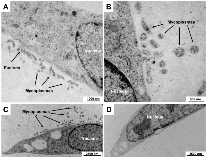
Figure 4.
Clearance of mycoplasma contamination in a rat cell culture as assessed by electron microcopy. A contaminated cell culture was treated with Plasmocin for 2 weeks. Shown are representative electron microscopic images of cells before (A–C) and after (D) treatment with the broad-spectrum anti-mycoplasma reagent showing that the mycoplasmas were successfully eliminated from the culture. Images were taken at original magnifications of 4646× to 27,800×. Please note the characteristic fusions of the mycoplasma membrane with the cytoplasmic membrane of the host cell in (A).

