Figure 2.
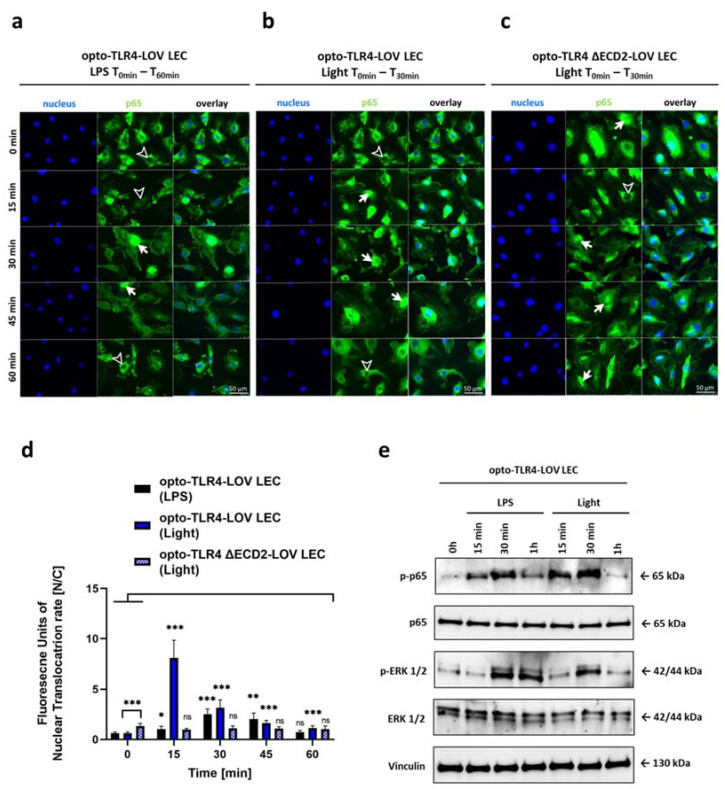
Spatiotemporal kinetics of p65. Intracellular localization of the p65 subunit of NF-κB was detected by immunofluorescence staining in opto-TLR4-LOV LECs treated with (a) LPS (100 ng/mL) or (b) blue light (470 nm) for 30 min or in (c) blue-light-treated opto-TLR4 ΔECD2-LOV. Then, p65 was stained using rabbit anti-p65 as the primary antibody and AF488-conjugated goat anti-rabbit IgG as the secondary antibody (green). Cell nuclei were stained using Hoechst 33342 (blue). Immunofluorescence images were acquired every 15 min for one hour. Arrows indicate prominent p65-NF-κB immunostaining in cell nuclei, while arrowheads display nuclei with deficient p65 labelling. Scale bar = 50 μm. (d) Quantitative evaluation of p65-NF-κB nuclear translocation. The mean ratio of relative fluorescence units of stained p65 in the nucleus to the cytoplasm ± standard deviation was graphically displayed in a bar chart (n = 6). Post-ANOVA, multiple comparisons relative to the control were performed using Šidák’s test (ns = not significant, * p < 0.05, ** p < 0.01, *** p < 0.001). (e) opto-TLR4-LOV LECs were continuously stimulated with LPS (100 ng/mL) or exposed to blue light (470 nm) for 1h. Western blots of whole-cell extracts were probed with antibodies against phospho-p65 (p-p65), p65, phospho-extracellular-signal-regulated kinase 1/2 (p-ERK1/2), ERK1/2, and vinculin as a loading control.

