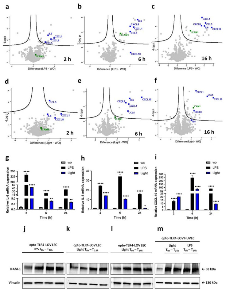
Figure 5.
Temporal proteomic and genomic expression analysis of pro-inflammatory mediators. Volcano plots show the regulation of over 1000 proteins in the supernatant of opto-TLR4-LOV LECs after 2 h, 6 h, and 16 h of (a–c) LPS (100 ng/mL) or (d–f) blue light (470 nm) stimulation compared to the untreated cells. The difference in LFQ protein abundance between ;reatment and control (wo) (x-axis) was plotted against its significance (-log p-value) (y-axis) (n = 4). Representative cytokines/chemokines (CXCL-1, CCL-5, IL-6, CXCL-8, and CXCL-10) (blue) and the typical adhesion molecule ICAM-1 (green) were annotated with their corresponding gene name. Relative (g) IL-6, (h) IL-8, and (i) CXCL-10 mRNA expression in opto-TLR4-LOV LECs 2 h, 6 h, and 24 h post-LPS (100 ng/mL) or blue light (470 nm) stimulation. The mRNA target gene expression levels were calculated according to the comparative CT method (2-ΔΔCT). Mean values ± standard deviation were graphically displayed in bar charts (n = 3). Post-ANOVA, multiple comparisons relative to the control were performed using Dunnett’s test (ns=not significant, ** p < 0.01, **** p < 0.0001). Opto-TLR4-LOV LECs were (j) continuously stimulated with LPS (100 ng/mL), (k) exposed to blue light (470 nm) for 30 min and then put into dark, or (l) continuously exposed to blue light (470 nm). (m) Opto-TLR4-LOV HUVECs were continuously treated with LPS (100 ng/mL) or illuminated with blue light (470 nm) for 24 h. Western blots of whole-cell extracts were probed with antibodies against ICAM-1 and vinculin as a loading control.

