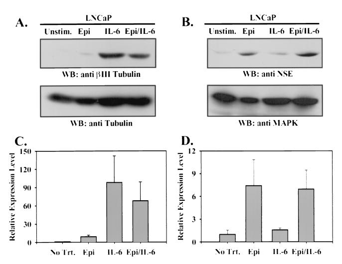
FIG. 3.
Epi and IL-6 induce expression of distinct neuronal markers. Immunoblots (WB) were prepared from 50 μg of whole-cell lysate of LNCaP cells cultured for 3 days in T-medium plus 5% FBS (Unstim.) or stimulated with Epi and IBMX (Epi), IL-6, or Epi, IBMX, and IL-6 (Epi/IL-6) as for Fig. 1 with anti-βIII tubulin antiserum (A, upper panel), total tubulin (A, lower panel), anti-NSE antiserum (B, upper panel), or anti-MAPK antiserum (B, lower panel). Expression of each marker was quantitated by densitometric scanning of the autoradiograms and expressed as the mean fold change in βIII tubulin expression relative to total tubulin expression (C) and the fold change in NSE expression relative to MAPK expression (D), normalized to the ratio in unstimulated cells ± SEM from three independent experiments.

