FIG. 7.
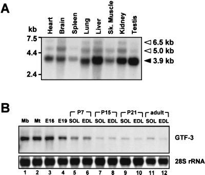
GTF3 expression in adult tissues and during muscle development. (A) Expression pattern of GTF3 in different mouse tissues. A commercial poly(A) blot containing 2 μg of mRNA from various adult mouse tissues per lane (mouse MTN; Clontech) was probed with a 32P-labeled full-length mouse GTF3 cDNA. The positions of RNA molecular weight markers are shown on the left. Filled arrowhead, position and size of the major GTF3 transcript; open arrowheads, additional bands that possibly represent incompletely processed or alternative GTF3 transcripts. Sk. muscle, skeletal muscle. (B) GTF3 expression is downregulated during muscle development. Total RNA from cultured myogenic cells and rat tissues (10 μg) was separated on a 1.5% formaldehyde-agarose gel and probed as described above. 28S rRNA hybridization signals are shown as a loading control. Total RNA from C2C12 myoblasts (lane 1) and myotubes (differentiated for 72 h; lane 2) were used to assess GTF3 expression during myogenic differentiation in vitro. RNA from whole hind limbs of E16 and E19 (excluding skin) rat embryos was used to approximate GTF3 expression during the fetal stage of appendicular muscle development (lanes 3 and 4). RNAs from postnatal stages of muscle development (from P7 to the adult; lanes 5 to 12) were used to determine GTF3 expression in maturating slow soleus (SOL) and fast EDL muscles.

