
FIG. 1.
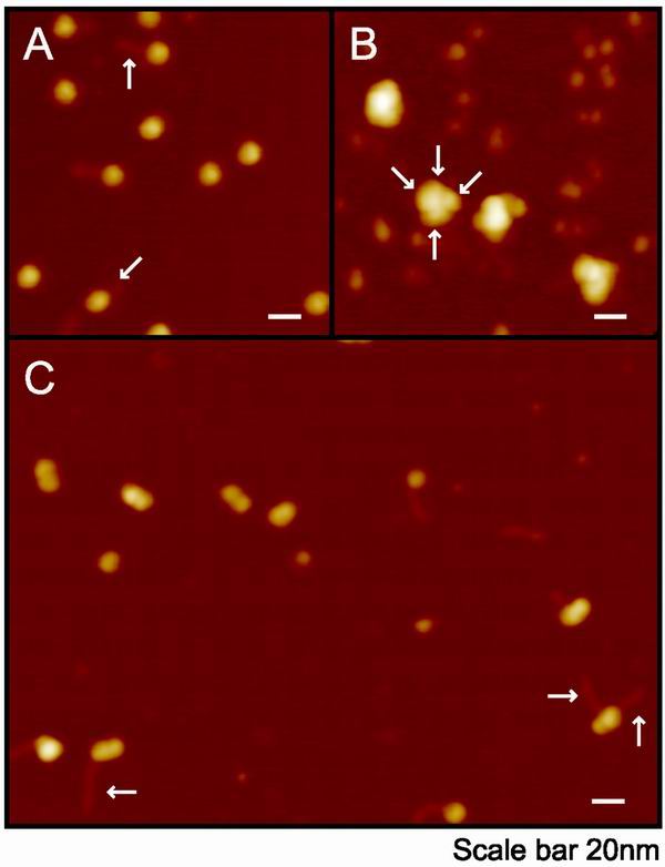
AFM images of SWI/SNF and altered dimers. Samples were fixed, deposited, and imaged with a nanotube tip as described in Materials and Methods. (A) Mononucleosomes on spermidine-treated mica. DNA tails, where visible, are indicated by arrows. (B) Gradient-purified hSWI/SNF on spermidine-treated mica. Multiple lobes are indicated by arrows. Small molecules are BSA from the gradient buffer. (C) hSWI/SNF-remodeled dimers on poly-l-lysine-treated mica. DNA tails are indicated by arrows.