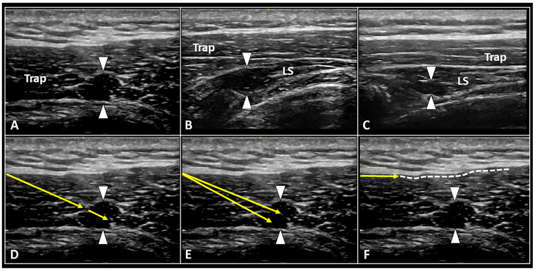
Figure 1.
Ultrasound images show MTrPs (white arrowheads) at the level of cervical spine located both within superficial muscles (A), e.g., upper fibers of trapezius (Trap), and deeper muscle planes (B,C), e.g., the levator scapulae (LS). Under real-time US guidance, back-and-forward movements of the needle (yellow arrow) to ‘release’ the nodule (D), fan-like dry needling of the trigger point (E), and hydro-dissection (F) of the histological interface between the muscle and the deep fascia (white dotted line) can be performed in a single multi-step procedure.

