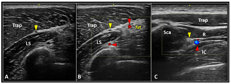
Figure 2.
Longitudinal US scan (A) clearly shows the anatomical location of the spinal accessory nerve (yellow arrowhead) within the interfascial space between the trapezius (Trap) and levator scapulae (LS) muscles. Accurately setting the color Doppler box, superficial and deep branches of the transverse cervical artery (red arrowheads) can be promptly identified to plan a safe intervention (B). Transverse ultrasound scan with color Doppler imaging (C) shows the dorsal scapular nerve (yellow arrowhead) and artery (red arrowhead) running inside the interfascial plane between the rhomboid (R) and intercostalis (IC) muscles medial to the medial edge of the scapula (Sca).

