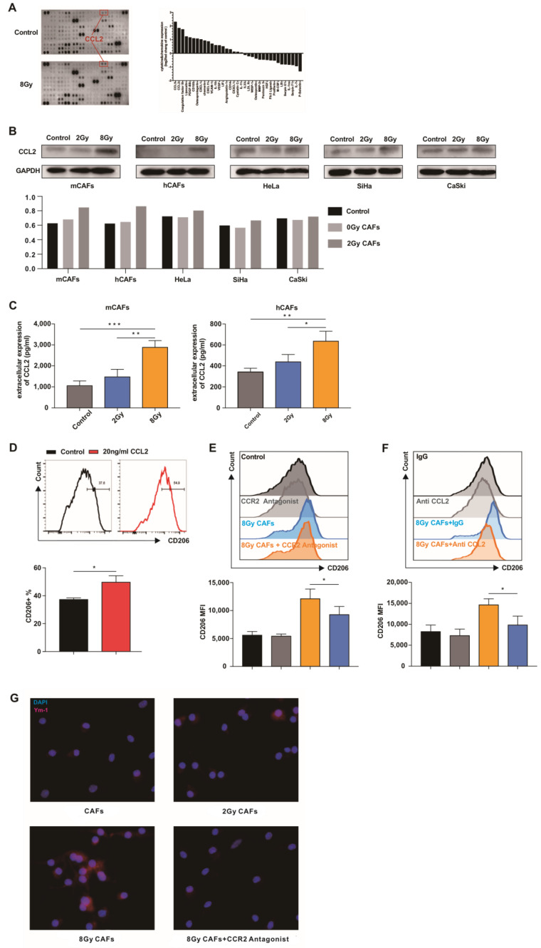
Figure 5.
CCL2 is critical for high-dose irradiated CAFs to promote M2 polarization. (A) mCAFs were exposed to 8 Gy radiation or not (control), and the supernatant of mCAFs was subjected to multiplex cytokine array analysis 24 h later. Left: a representative blot. Right: quantification of dot intensity of significantly changed cytokines. (B) Cervical cancer cell lines and CAFs were irradiated with the indicated doses. After 24 h, CCL2 expression was analyzed by Western blot analysis. (C) mCAFs and hCAFs were exposed to the indicated doses of radiation. After 24 h, the supernatant was subjected to ELISA. Data are shown as mean ± SD from three independent experiments. The data were analyzed using one-way ANOVA. * p < 0.05, ** p < 0.01, *** p < 0.001. (D) BMDMs were treated with 20 ng/mL CCL2 for 24 h and then subjected to flow cytometry analysis. Data are shown as mean ± SD from three independent experiments. Differences between groups were analyzed using unpaired Student’s t-test. * p < 0.05. (E) M0 BMDMs treated with 10 nM CCR2 antagonist (INCB3344) were co-cultured with irradiated or non-irradiated mCAFs for 3 days. Then, the cells were subjected to flow cytometry analysis to evaluate CD206 expression. Data are shown as mean ± SD from three independent experiments. * p < 0.05. (F) Anti-CCL2 neutralizing antibody (20 μg/mL) was added to a co-culture system consisting of 8 Gy irradiated mCAFs and M0 BMDMs. The BMDMs were subjected to flow cytometry analysis after 3 days of co-cultivation. Data are shown as mean ± SD from three independent experiments. The data were analyzed using one-way ANOVA. * p < 0.05. (G) M0 BMDMs were co-cultured with mCAFs exposed to the indicated doses of radiation for 3 days and 10 nM CCR2 antagonist (INCB3344) was added into the 8 Gy irradiated mCAF co-culture system. Immunofluorescence analysis of the expression of Ym-1 (red) in BMDMs. The whole western blots are shown in File S1.

