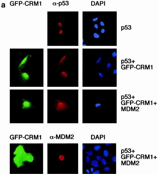
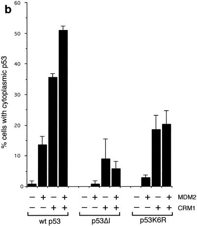
FIG. 7.
(a) Subcellular localization of p53, MDM2, and GFP-CRM1 following transfection into U2OS cells. Costaining with DAPI shows localization of the nucleus. (b) Graph showing the proportion of cells expressing cytoplasmic p53 following transfection of wild-type p53, p53ΔI, or p53K6R, with GFP-CRM1 and MDM2 in U2OS cells. Note the reduced sensitivity of detection of cytoplasmic localization of untagged p53 compared to GFP-p53 shown in Fig. 5b and 6b.


