
FIG. 5.
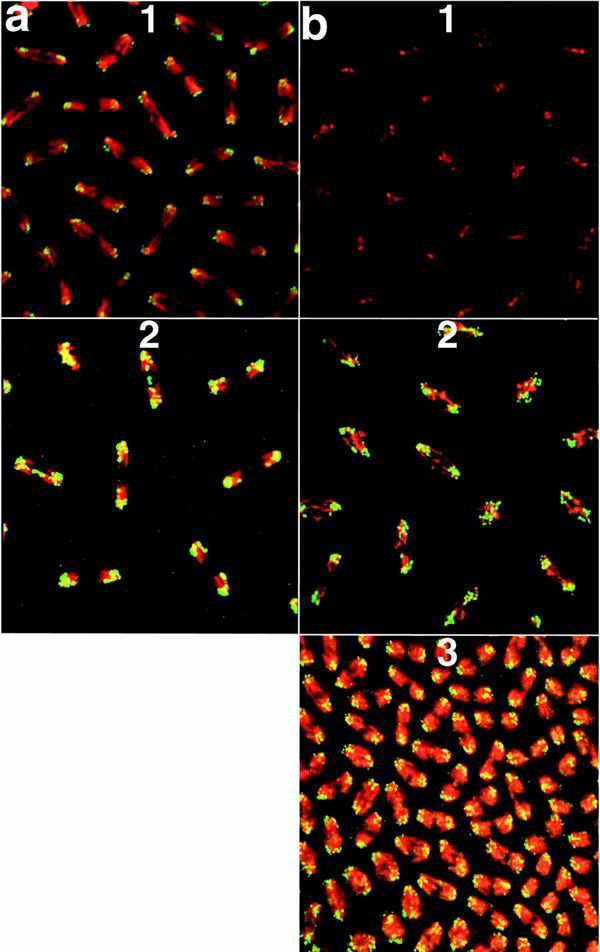
Association of GAGA-519 and GAGA-581 with centric heterochromatin. DNA is in red, and protein is in green. Chromosomes are shown in anaphase, at the time they are being separated by microtubules. Centromeres are pointing outward, and telomeres are oriented inward. All embryos are at the syncytial blastoderm stage (stage 4 [36]) of embryogenesis but have undergone different numbers of nuclear divisions. Embryos were stained with the GAGA-519-specific antibody (a) or with the GAGA-581-specific antibody (b). DNA was visualized with TOTO-1. The genotypes of embryos were as follows: a1 and b1, wild type; a2, 4Xhsp83: GAGA-519; b2, Xhsp83: GAGA-581; b3, 4Xhsp83GAGA-581; Trl109.2/TrlR67.