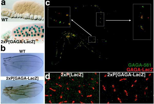
FIG. 7.
Localization of the hsp83:GAGA-LacZ transgene. (a) Salivary glands from wild-type (WT) and 2Xhsp83:GAGA-LacZ larvae stained with X-Gal. Line #8 of hsp83:GAGA-LacZ was used. Other lines showed similar staining patterns and intensities (data not shown). (b) Wing defects associated with GAGA-LacZ. Flies were raised at 29°C, and wings were dissected off and mounted in Hoyer's mountant. (c) Imperfect colocalization of GAGA-581 (green) and GAGA-LacZ (red). The overlap is yellow. Polytene chromosomes were double stained using antibodies against GAGA-581 and LacZ, as described in Materials and Methods. (d) Association of GAGA-LacZ with centric heterochromatin. DNA is in red, and protein is in green. As in Fig. 4, chromosomes are shown in anaphase, at the time they are being separated by microtubules. Centromeres are pointing outward, and telomeres are oriented inward. Embryos were stained with an antibody against LacZ; DNA was visualized with TOTO-1 (see Materials and Methods).

