Figure 3.
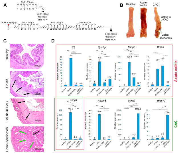
Morphological changes and expression levels of key genes identified by the bioinformatics analysis in the colon tissues of healthy mice and mice with acute colitis and colitis-associated cancer (CAC). (A) Experimental setup. Acute colitis was induced by the administration of 2.5% dextran sulfate sodium (DSS) solution in drinking water for 7 days followed by a 3 day recovery (upper panel) (n = 10). CAC was induced in mice by single intraperitoneal (i.p.) injection of azoxymethane (AOM) 1 week before DSS administration. Mice were then exposed to 3 consecutive cycles of 1.5% DSS instillations for 7 days followed by 2 weeks recovery (lower panel) (n = 10). At the end of the experiment, the colons were collected for subsequent histological analysis and qRT-PCR. (B,C) Gross morphology (B) and histology (C) of the colon tissue of healthy mice and mice with acute colitis and CAC. Hematoxylin and eosin staining. Original magnification ×100. Black arrows indicate inflammatory infiltration. Green arrows indicate adenomatous transformation of colon epithelium. (D) Expression levels of identified genes in the colon tissues measured by qRT-PCR. HPRT was used as a reference gene. Healthy—healthy colon tissue, colitis—colon tissue with acute colitis, colitis in CAC—colon tissue with chronic colitis adjacent to adenomas. The data are expressed as the mean ± SD (n = 6). * p < 0.05, ** p < 0.01, *** p < 0.001.

