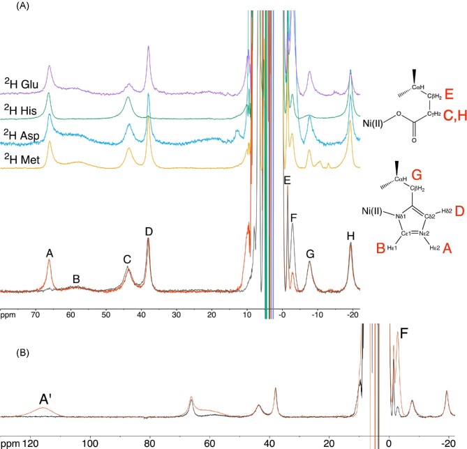
Fig. 8.
1H NMR spectra (400 MHz) of (A) Ni,Zn-HypA in H2O (red) and D2O (gray), together with the 1H NMR spectra of the corresponding selectively deuterated forms (2H Met in orange, 2H Asp in blue, 2H His in green and 2H Glu in purple). The assignment of signals belonging to His2 is shown. Panel B shows the 1H NMR spectrum of Ni,Zn-HypA in the presence (red) and absence (black) of excess Ni.

