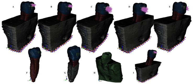
Figure 1.
Mesh model: (A) 2nd lower right premolar model with 4 mm bone loss and applied vector for extrusion; (B) 2nd lower right premolar model with 4 mm bone loss and applied vector for intrusion; (C) 2nd lower right premolar model with 4 mm bone loss and applied vector for rotation; (D) 2nd lower right premolar model with 4 mm bone loss and applied vector for tipping; (E) 2nd lower right premolar model with 4 mm bone loss and applied vector for translation, (F) tooth model; (G) tooth model with 4 mm reduced PDL; (H) no bone loss 3D model; (I) 2nd lower right premolar model with 8 mm bone loss and applied vector for extrusion.

