Figure 5.
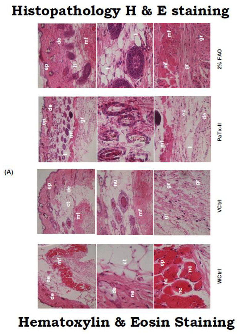
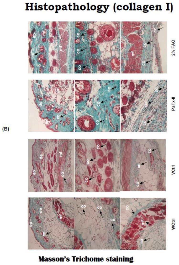
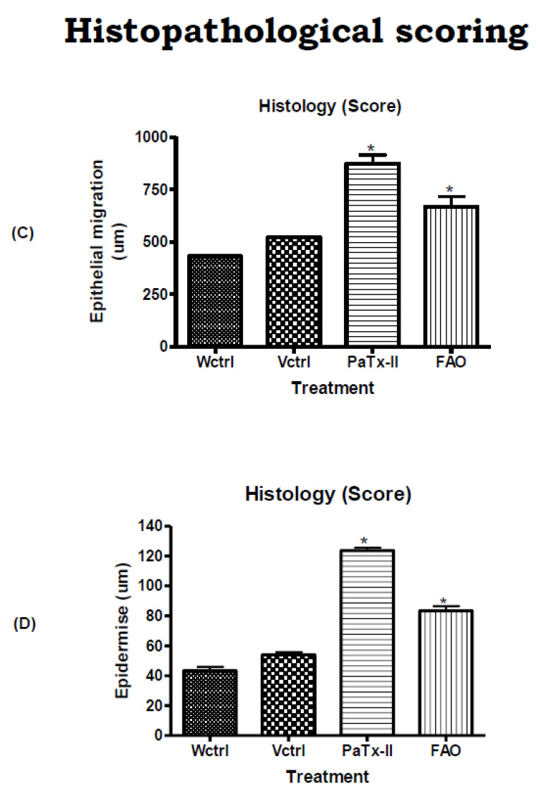
(A) Pathological assessment of wounds before and after treatment was evaluated under the light microscope. Group (I): WCtrl mice show redness and inflammation. Group (II): Among VCtrl mice, there was necrosis (nc) and abundant inflammatory infiltrates, changes in the muscle fibers (mf) and almost complete destruction of the mf cells after injury on day 14. Group (III): PaTx-II locally applied to mice exerted significant healing and clearing of bacteria after 14 days. Group (IV): Two percent FAO-treated mice healed more slowly than the PaTx-II treatment group. Abbreviations: ep-epidermis, de-dermis, li-leukocyte infiltration, mf-muscle fiber, nu-neutrophils, gr-granules, ne-necrosis (original magnification: ×20–×100). (B) MT staining detection of collagen deposition in skin tissues of different wound healing groups before and after treatment. G (I)—WCtrl and G(II)—VCtrl collagen was not detected in either group up 14 days post-wounding. G (III): PaTx-II-treated mice show abundant expression of collagen at 14 days after local application. G (IV): Two precent FAO-treated mice also equally expressed collagen for complete healing (original magnification: ×20-×100). (C) Epithelial migration from the wound edge increases the thickness of the epithelial layer 14 days post wounding. (D) Epidermal migration from both edges of the wound was measured with an ocular micrometer. Three measurements were used for each wound (n = 3); values are expressed as mean of epithelial migration ± SD thickness of epithelium. Both treatments show a significant difference (* p < 0.05) from controls (WCtrl and VCtrl), and there was also a significant difference (* p < 0.05) between PaTx-II and FAO-treated mice. ‘co’ and ‘ct’ on images represent controls.


