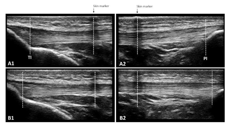
Figure 3.
The overlapping images technique enables the measurement of patellar tendon length at rest (A1 + A2) and in increments of 10% force up to maximum voluntary isometric contraction (B1 + B2) when the entire length cannot be captured in a single frame due to the limited size of the ultrasound probe. A skin marker (adhesive tape) that creates a hypoechoic shadow is used to define the measurement bounds: the length from TI to the center of the skin marker in A1 and the length from the center of the skin marker to PI in A2 are added. The same procedure is repeated for each 10% increase in force, leading to B1 and B2.

