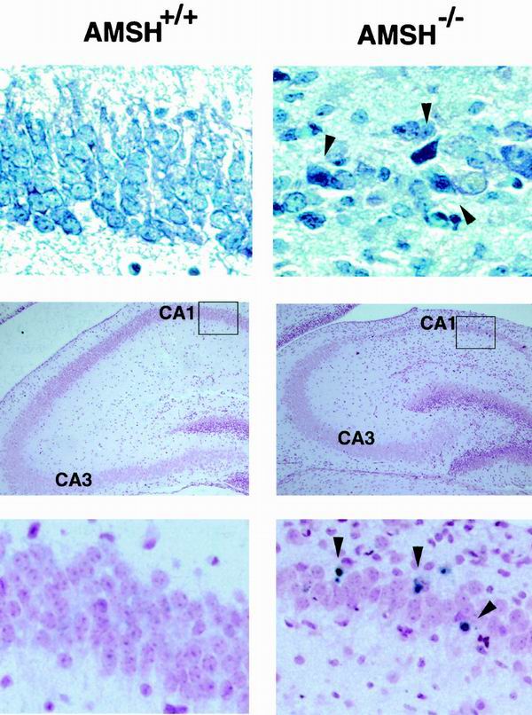
FIG. 5.
Appotosis of the AMSH-deficient hippocampal neurons in vivo. The top two panels show toluidine blue staining of anterior coronal hippocampus sections; the four lower panels show TUNEL staining of anterior coronal hippocampus sections. The figure shows AMSH+/+ mice (left panels) and AMSH−/− mice (right panels) at 6 days old. The bottom two panels represent the magnified views of the areas in the boxes as shown in the middle two panels (left and right, respectively). Arrowheads in the top right and bottom right panels indicate pyknotic and TUNEL-positive cells, respectively. Magnifications, approximately ×300 (top and bottom panels) and approximately ×60 (middle panels).

