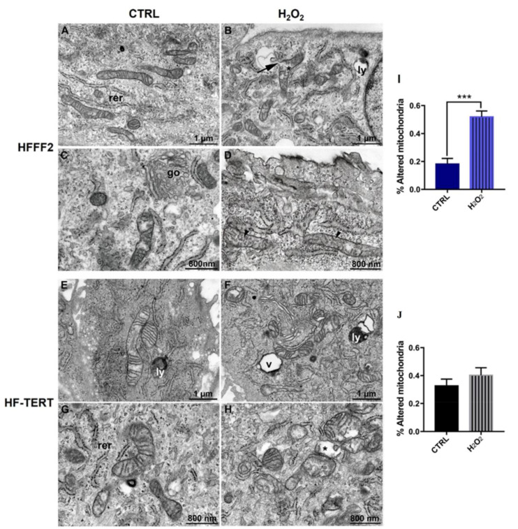
Figure 7.
FIB/SEM micrographs illustrating mitochondrial morphology in HFFF2 (A–D) and HF-TERT (E–H) cells, treated and untreated with H2O2. In untreated cells, mitochondria display a typically elongated shape and numerous parallel-oriented cristae. Following treatment, mitochondrial swelling (asterisks), with matrix expansion and cristae dysmorphic features, including numerical reduction, spatial disorganization and/or fragmentation (arrowheads) are observed. Rupture of the outer membrane is occasionally found (arrow). The diagrams report mitochondria alteration in untreated and treated (I) HFFF2 cells and (J) HF-TERT cells after 3-hour recovery. Data represent mean ± SEM. *** p < 0.001; by Student’s t-test. Acronyms: (go) Golgi complex, (ly) lysosome, (rer) rough endoplasmic reticulum, (v) vacuole.

