Figure 2.
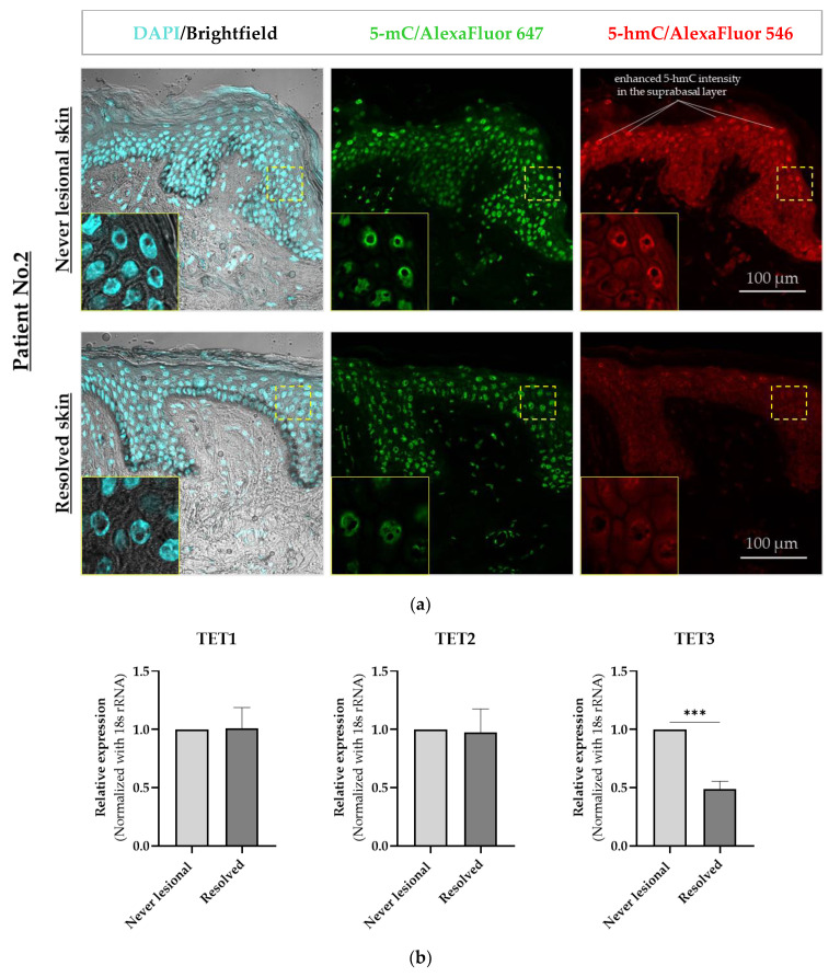
Confocal microscopy immunofluorescence analysis of 5-mC and 5-hmC in never-lesional vs. resolved skin. (a) Representative images are shown (n = 4). Colocalization immunofluorescence staining of 5-mC (green) and 5-hmC (red). Closeup images show yellow-dotted regions at higher magnification. Scale bars are 100 μm. (b) Real-time RT-PCR of TET1, TET2, and TET3 in resolved epidermis vs. never-lesional epidermis. An unpaired two-tailed t-test was performed, *** p < 0.001. Data = mean ± SEM of biological replicates (n = 3). Abbreviations: 5-mC, 5-methylcytosine; 5-hmC, 5-hydroxymethylcytosine; TET1, ten-eleven translocation (TET)1; TET2, ten-eleven translocation (TET)2; TET3, ten-eleven translocation (TET)3.

