Figure 3.
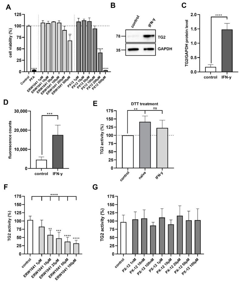
Evaluation of TG2 inhibition on confluent Caco-2 cell monolayers. (A) Cell viability of Caco-2 cells was tested after 24 h treatment with ERW1041 and PX12 at the indicated concentrations. n = 3 with three technical replicates per experiment. (B) Protein expression of TG2 in Caco-2 cells was analyzed by Western blotting after treatment IFN-γ. GAPDH served as loading control. (C) Relative quantities were normalized to GAPDH levels in whole cell lysates. IFN-γ stimulation significantly increased TG2 levels in differentiated Caco-2 cells (n = 4). (D) TG2 activity in confluent Caco-2 cell monolayers was investigated by fluorometry after stimulation with IFN-γ (1000 IU/mL) for 48 h. IFN-γ treatment significantly increased TG2 activity on the cell surface. n = 6 with three technical replicates per experiment. (E) Naïve and IFN-γ-stimulated Caco-2 cells were treated with 1 mM DTT in the presence of 5BP. Crosslinking of 5BP was evaluated by fluorometry. In non-stimulated Caco-2 cells, DTT treatment significantly increased TG2 activity, but not in IFN-γ-stimulated Caco-2 cells. n = 4 with three technical replicates per experiment. (F) Caco-2 cells were incubated with 5BP in the presence of different amounts of ERW1041 for 3 h. TG2-mediated crosslinking was quantified by fluorometry. ERW1041 inhibited extracellular TG2 in a dose-dependent manner. n = 4 with three technical replicates per experiment. (G) PX-12 did not inhibit TG2 activity on confluent monolayers of Caco-2 cells at the tested concentrations. n = 4 with three technical replicates per experiment. All data are shown as mean ± SD. Statistical significance was tested with Student’s t-test (with Welch correction where appropriate) and one-way ANOVA with Dunnett’s multiple comparison test (E), ** p < 0.01, *** p < 0.001, **** p < 0.0001; ns = not significant.

