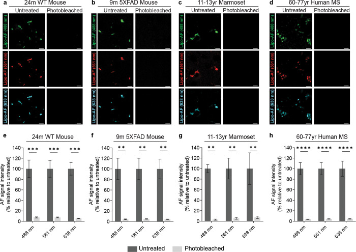
Fig. 5. Photobleaching quenches lipo-AF signal in mouse, marmoset and human brain tissue.
a-d Representative images of lipo-AF upon excitation with a 488 nm, 561 nm or 638 nm laser line in untreated (left column) or photobleached (right column) samples from 24-month-old mouse cortex (a), 9-month-old 5XFAD mouse cortex (b), 11–13 year-old marmoset cortex (c), and 60–77 year-old human cortex (d). Scale bars = 5 μm. e-h Quantification of lipo-AF signal intensity before and after photobleaching for each species. Data represented as a mean ± SEM, n = 3 biological replicates per species. Two-way ANOVA with Šídák’s multiple comparisons test (e, F = 114.7, df = 1; d, F = 69.41, df = 1; f, F = 59.30, df = 1; h, F = 171.3, df = 1;); **p<0.01, ***p<0.001****p<0.0001.

