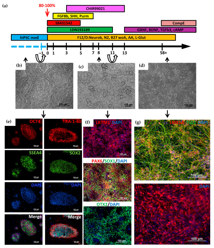
Figure 1.
Directed differentiation of induced pluripotent stem cells (iPSCs) into dopaminergic (DA) neurons. (a) Scheme of iPSCs’ differentiation into DA neurons. Cell passaging was performed on –1st and 11th days of differentiation; (b) Morphology of iPSCs cultured on MEF; (c) Monolayer of neuroectodermal cells on day 8 of differentiation; (d) Morphology of terminally differentiated neurons. Phase contrast (b–d); (e) Immunofluorescent staining of iPSCs for pluripotency markers: transcription factors OCT4 (red signal), SOX1 (green signal) and surface markers TRA-1-60 (red signal), SSEA4 (green signal); (f) Immunofluorescence staining for DA neuron progenitor marker LRTM1 (red signal) and early neuroectoderm markers: PAX6 (red signal), OTX2 (green signal), SOX1 (green signal); (g) Immunofluorescent staining for markers of terminally differentiated DA neurons: tyrosine hydroxylase (TH, red signal) and LMX1A (red signal). Nuclei are stained with DAPI (blue signal). Scale bar: 100 µm.

