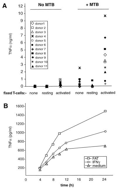
FIG. 1.
FAT synergize with M. tuberculosis for enhanced TNF-α production. (A) Primary monocytes were cultured in the absence or presence of M. tuberculosis (MTB; 1 bacterium per monocyte) as indicated. Some of these cultures also received 1 fixed HUT T cell (resting or activated) per monocyte, as indicated, at the time of infection. Twenty-four hours later, supernatants were collected and assayed for TNF-α by ELISA. Each symbol represents 1 of 11 donors and indicates the mean of triplicate cultures from an individual donor. M. tuberculosis and FAT individually stimulated (P < 0.001) TNF-α production compared to unstimulated monocytes. However, TNF-α production from cultures receiving both FAT and M. tuberculosis was significantly higher (P < 0.001) compared to cultures receiving either stimulus alone. Time course of TNF-α release is shown in panel B. Monocytes were stimulated as above with M. tuberculosis in the absence or presence of either FAT or IFN-γ (100 U/ml) as shown. Supernatants were harvested at the indicated time points and assayed for immunoreactive TNF-α. Results shown are from one representative of three experiments.

