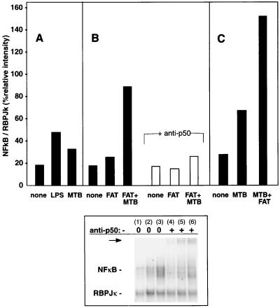
FIG. 5.
NF-κB activation by M. tuberculosis in the absence or presence of FAT membranes. Monocytes were stimulated with LPS (1 μg/ml) or M. tuberculosis (MTB; 3 γ-irradiated bacteria per monocyte) and/or FAT membranes (the equivalent of 3 T cells per monocyte) as indicated for 90 min before nuclear proteins were extracted and assayed by EMSA. Results shown are the percentage of radiolabeled probe binding to NF-κB relative to a constitutive internal control (RBPJκ) from one representative of two experiments for each of A, B, and C (solid bars). In addition, the identity of the NF-κB was confirmed by gel retardation with anti-NF-κB p50 antiserum (B; open bars; arrow, bottom panel). The lanes in the lower panel correspond to the bars shown in B as follows: 1 and 4, unstimulated; 2 and 5, FAT; 3 and 6, FAT plus M. tuberculosis; 1 to 3, no antibody; 4 to 6, plus anti-p50.

