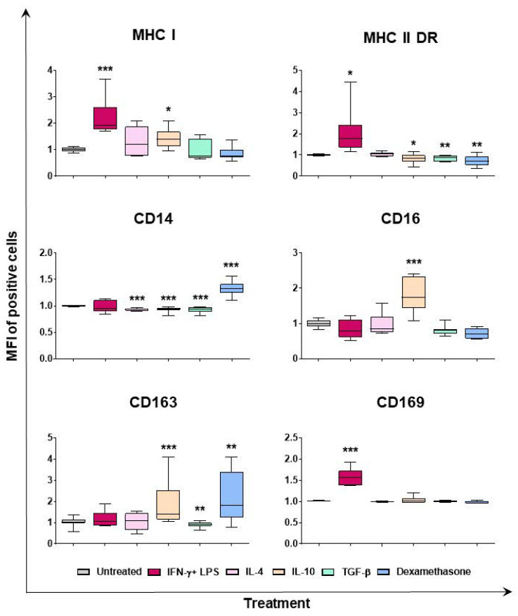
Figure 2.
Effect of diverse polarizing factors on porcine moMΦ surface marker expressions (mean of fluorescence intensity). Porcine moMΦ were left untreated, or they were stimulated with diverse polarizing factors: IFN-γ + LPS (both at 100 ng/mL), IL-4 (20 ng/mL), IL-10 (20 ng/mL), TGF-β (20 ng/mL), or dexamethasone (20 ng/mL). Then, 24 h post-stimulation, flow cytometry was employed to determine the expression of several surface markers: MHC I, MHC II DR, CD14, CD16, CD163, and CD169. Mean fluorescence intensity (MFI) of positive cells was evaluated, and MFI data are expressed as fold change relative to the un-activated condition (moMΦ). Data from three independent experiments utilizing different blood donors are presented. Data are displayed as box-and-whisker plots, showing the median and interquartile range (boxes) and minimum and maximum values (whiskers). Values of treated macrophages were compared to the untreated control (moMΦ), using an unpaired t-test of a Mann–Whitney test. *** p < 0.001, ** p < 0.01, and * p < 0.05.

