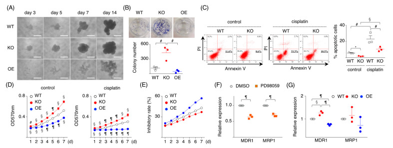
Figure 3.
SPRED2 as a regulator of cancer cell stemness. (A) WT, SPRED2-KO, and SPRED2-OE cells (1000 cells) were cultured in 96-well ultra-low attachment plates for 14 days. Representative photos are shown. Scale bars: 100 μm. (B) Spherical colonies were dissociated, and 1 × 10⁴ cells were planted into a 6-well plate, and cultured for an additional 14 days. Upper panel, representative photos are shown. Lower panel, colony numbers in 6-well plates were counted (n = 3, each). (C) WT and SPRED2-KO cells (106 cells) were cultured with or without cisplatin (10 μg/mL) for 24 h. Left panel, cells were stained with FITC-Annexin V and PI, and the percentage of apoptotic cells was analyzed by flowcytometry. Right panel, the %apoptotic cells were evaluated (n = 3, each). (D) WT, SPRED2-KO, and SPRED2-OE cells were treated with or without cisplatin (10 μg/mL) for 24 h. Cell proliferation was evaluated by MTT assay (n = 3, each). (E) Percent inhibitory rate was calculated by (1-cisplatin: A570/control: A570) ×100. (F) WT cells were treated with 20 μM PD98059 or vehicle (DMSO) for 24 h. MDR1 and MRP1 mRNA expressions in cell extracts were examined by RT-qPCR (n = 3, each). (G) The mRNA expressions of MDR1 and MRP1 in WT, SPRED2-KO and SPRED2-OE cell lysates were evaluated by RT-qPCR (n = 3, each). # p < 0.05, § p < 0.01, ¶ p < 0.001, * p < 0.0001, two-tailed unpaired t test.

