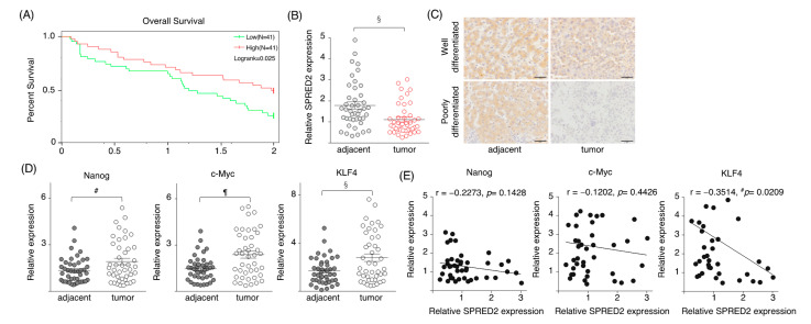
Figure 6.
SPRED2 expression in HCC tissues. (A) Among 371 HCC cases from TCGA database, 82 cases that could be followed for 2 years, including those who died, were selected. The SPRED2 expression of these 82 cases was dichotomized, and a survival curve was drawn with the high 50% as SPRED2-high and the low 50% as SPRED2-low. (B–E) Data from our 40 HCC patients. (B) The expression levels of SPRED2 mRNA. (C) Representative photos of SPRED2 immunohistochemistry from well and poorly differentiated HCC tissues. Scale bars: 50 μm. (D,E) Expressions of pluripotency factors in HCC tissues. mRNA was extracted from paraffin block. (D) The expression levels of Nanog, c-Myc and KLF4 were measured by RT-qPCR. (E) The relation between Nanog/c-Myc/Klf4 and SPRED2 was analyzed. # p < 0.05, § p < 0.01, ¶ p < 0.001, two-tailed unpaired t test.

