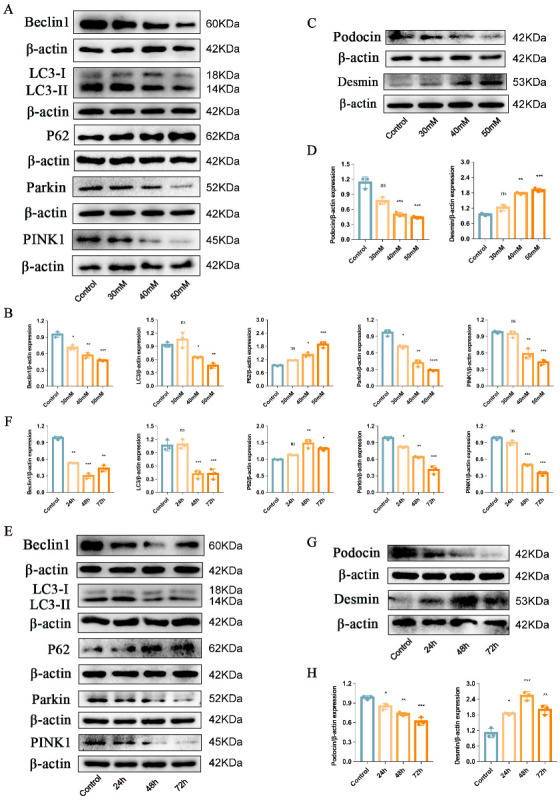
Figure 1.

Podocyte injury and PINK1/Parkin-mediated mitophagy inhibition induced by HG. (A,B) Representative Western blot analysis of Beclin1, LC3II/LC3I ratio, P62, Parkin, and PINK1 in MPC5 treated with different concentrations of glucose. (C,D) Representative Western blot analysis of Podocin and Desmin in MPC5 treated with different concentrations of glucose. (E,F) Representative Western blot analysis of Beclin1, LC3II/LC3I ratio, P62, Parkin, and PINK1 in MPC5 treated with HG for 24 h, 48 h, and 72 h. (G,H) Representative Western blot analysis of Podocin and Desmin in MPC5 treated with HG for 24 h, 48 h, and 72 h. n = 3 for (A–H). β-actin was used as loading control. * p < 0.05, ** p < 0.01, *** p < 0.001, and **** p < 0.0001 vs. control; ns = not significant.
