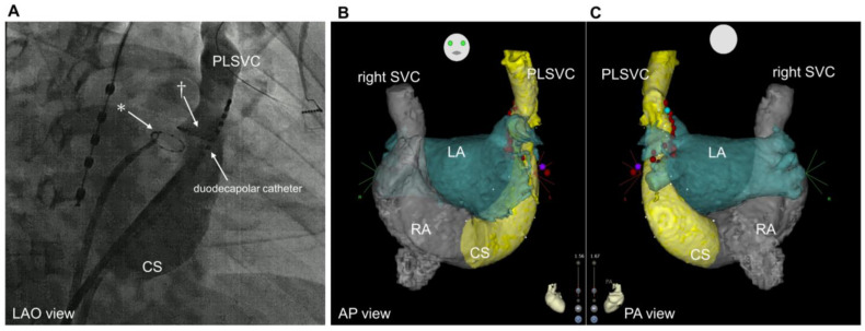
Figure 3.
Fluoroscopic images and computed tomography. (A): Coronary sinus venography performed with a long sheath placed within the dilated coronary sinus (LAO view). * Circular mapping catheter placed in the left inferior pulmonary vein † Circular mapping catheter placed in the PLSVC. (B): Preprocedural CT imaging. anteroposterior view. (C): Posteroanterior view. CS, coronary sinus; PLSVC, persistent left vena cava; LAO, left anterior oblique; SVC, superior vena cava; LA, left atrium; RA, right atrium; CT, computed tomography.

