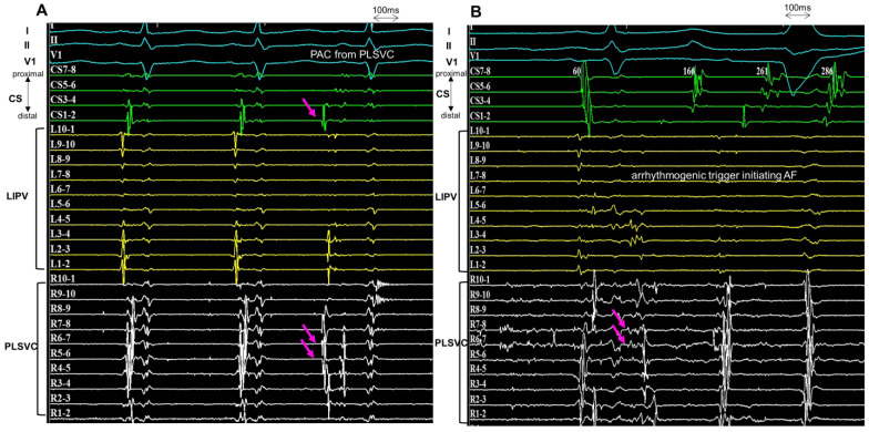
Figure 4.
Arrhythmogenic trigger from PLSVC. (A): The circular mapping catheters are positioned within the LIPV and PLSVC. During the first two sinus beats, the two components (left atrial far-field and PLSVC potential) are fused. During the ectopic rhythm (the third beat), the sharp potential of the PLSVC precedes the surface ECG by 60 ms. The atrial activation sequence is from distal to proximal on the CS during the PAC. (B): During AF initiation, the sharp potential of the PLSVC precedes the PAC, and the PAC arising from the PLSVC initiates the AF. CS, coronary sinus; PLSVC, persistent left vena cava; LIPV, left inferior pulmonary vein; PAC, premature atrial contraction; AF, atrial fibrillation.

