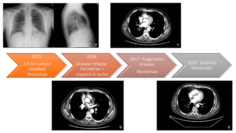
Figure 1.
The therapy response timeline from 2015 to today. (a) Chest X-ray (Thoravision). Examination performed with digital technologies in the two orthogonal projections. The parenchymal opacity already indicated on the right and better evident in the previous TC of 3/3/2015 is confirmed. Disventilatory streak in the left basal site. (b) Examination performed before and after intravenous administration of organ-iodized contrast medium place comparison with the previous PET-CT investigation carried out on 29 July 2016. Compared to the previous one, there is a documented increase in the size of the newly formed tissue located in the right hilar seat (current dimensions 27 × 25 mm vs. 20 × 11 mm of the previous exam). The findings in the lungs are unchanged, the thickening area previously described at the level of the LID. No pleural effusion flaps. (c) Examination carried out before and after administration of organ-iodized MDC by IV route, compared with previous PET-CT carried out on 5 July 2018. There are no areas of pathological enhancement in the sub- and supratentorial sites. The axial ventricular system showed dimensions within the limits of the norm. The results of the surgery known in the anamnesis in the thoracic area on the right have been confirmed. The millimetric nodular formation already evident at the lower lobe appears unchanged right in the previous aforementioned signs of emphysema in both parenchymal areas. No pleural effusion flaps have been observed. (d) Neck-thorax: Nodular thyroid isthmic formation requiring monitoring remains unchanged instrumental with ultrasound examination. The results of surgery on the upper-right lobe have been confirmed. The share of tissue in the right hilar seat and the solid nodular formation (4 mm) correspond to the LID. No pleural or pericardial effusion. Sub-centimetric lymph node formations in the hilar and mediastinal region were stable in size.

