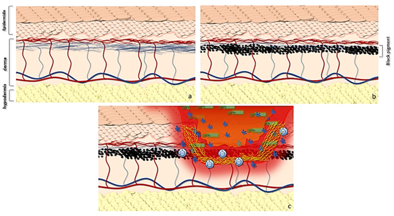
Figure 4.
Graphic abstract showing the possible evolution of gram+ skin infections in tattooed patients with immunotherapy. (a) Normal skin anatomy and trophism of epidermis. (b) The use of the pigment leads both to a local microtrauma and to the deposit of pigment in the superficial portion of the dermis with processes of inflammation at the time of tattooing and subsequent inflammation. At the same time, the pigment, together with microtrauma fibrosis, reduces the diffusion of transudate from the dermis to the epidermis, also decreasing the superficial trophism. (c) This determines a reduced barrier action of the epidermis, which supports Ruocco’s hypothesis. In LMR, infections can lead to a local destructive process evolving in phlegmon.

