Abstract
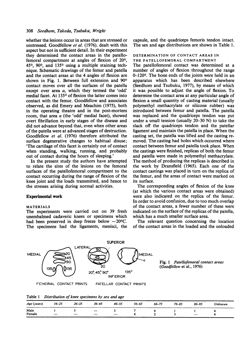
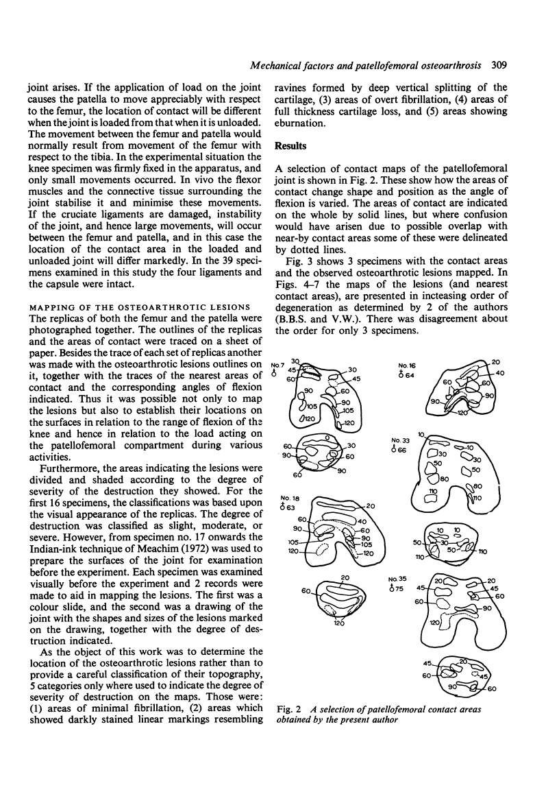
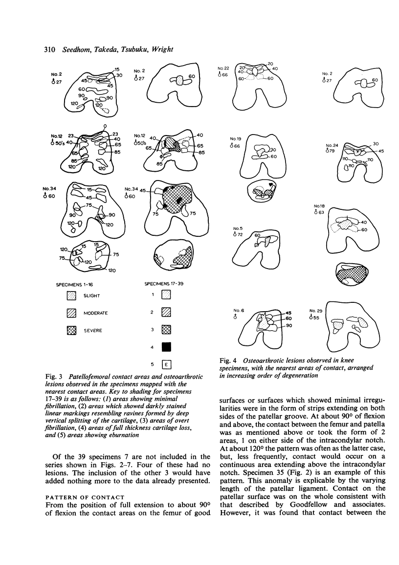
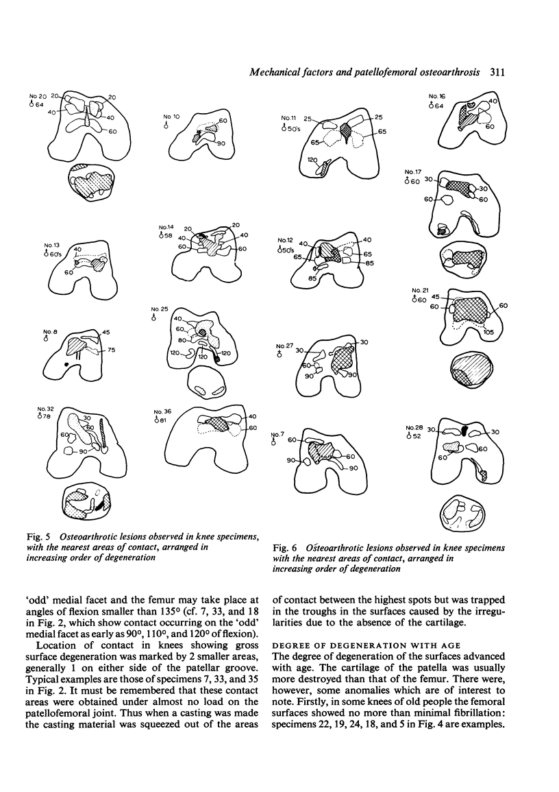
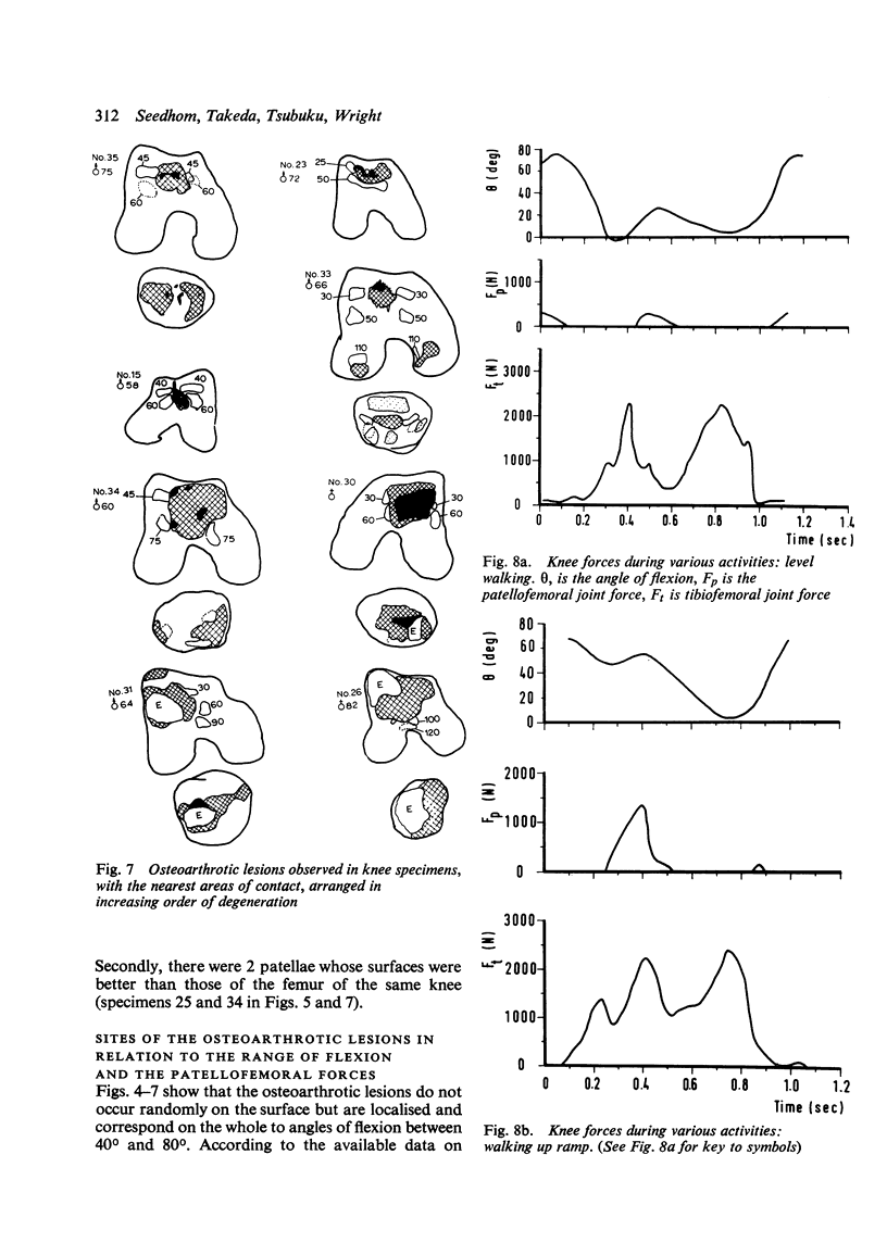
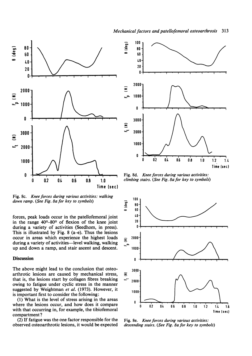
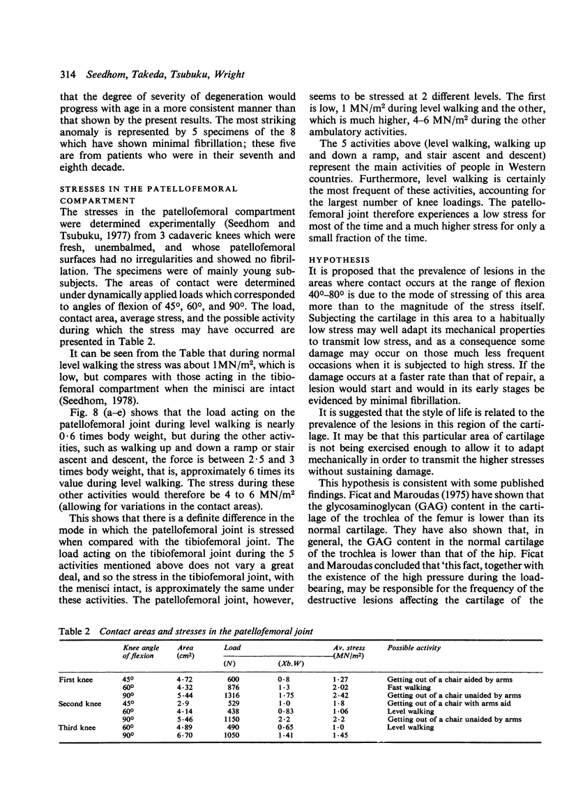
This paper investigates the possible role of mechanical stress in the development of the osteoarthrotic lesions frequently observed in the patellofemoral compartment of the knee joint. First the location of these destructive lesions was determined by studying the location and pattern of contact in the patellofemoral joint. The study was carried out on 39 cadaveric knees for the range of flexion 0 degrees -120 degrees. It was shown that the lesions were localised to the areas corresponding to the range of flexion 40 degrees -80 degrees. These areas have been shown to be subjected to a low stress for most of the time and to a much higher stress for only part of the time. This mode of stressing this area of the cartilage is a consequence of the style of life of the average Western man in which the most predominant activity is level walking, during which the load and in turn the stress are much lower than they are during other ambulatory activities such as ramp and stair ascent and descent. The same area of the cartilage seems to be subject to a similar mode of stress during sedentary occupations. It is suggested that this mode of stressing the cartilage conditions it chemically, and hence mechanically, to transmit low stresses, so that when the much less frequent but higher stresses are applied it cannot transmit them without sustaining some damage.
Full text
PDF
Selected References
These references are in PubMed. This may not be the complete list of references from this article.
- Dransfield B. S. The laboratory technique for the construction of a skull using cold cure acrylic. Dent Tech. 1965 Oct;18(10):201–206. [PubMed] [Google Scholar]
- Emery I. H., Meachim G. Surface morphology and topography of patello-femoral cartilage fibrillation in Liverpool necropsies. J Anat. 1973 Oct;116(Pt 1):103–120. [PMC free article] [PubMed] [Google Scholar]
- Ficat C., Maroudas A. Cartilage of the patella. Topographical variation of glycosaminoglycan content in normal and fibrillated tissue. Ann Rheum Dis. 1975 Dec;34(6):515–519. doi: 10.1136/ard.34.6.515. [DOI] [PMC free article] [PubMed] [Google Scholar]
- Goodfellow J. W., Bullough P. G. The pattern of ageing of the articular cartilage of the elbow joint. J Bone Joint Surg Br. 1967 Feb;49(1):175–181. [PubMed] [Google Scholar]
- Goodfellow J., Hungerford D. S., Zindel M. Patello-femoral joint mechanics and pathology. 1. Functional anatomy of the patello-femoral joint. J Bone Joint Surg Br. 1976 Aug;58(3):287–290. doi: 10.1302/0301-620X.58B3.956243. [DOI] [PubMed] [Google Scholar]
- Kempson G. E., Muir H., Swanson S. A., Freeman M. A. Correlations between stiffness and the chemical constituents of cartilage on the human femoral head. Biochim Biophys Acta. 1970 Jul 21;215(1):70–77. doi: 10.1016/0304-4165(70)90388-0. [DOI] [PubMed] [Google Scholar]
- LINN F. C., SOKOLOFF L. MOVEMENT AND COMPOSITION OF INTERSTITIAL FLUID OF CARTILAGE. Arthritis Rheum. 1965 Aug;8:481–494. doi: 10.1002/art.1780080402. [DOI] [PubMed] [Google Scholar]
- Meachim G., Emery I. H. Quantitative aspects of patello-femoral cartilage fibrillation in Liverpool necropsies. Ann Rheum Dis. 1974 Jan;33(1):39–47. doi: 10.1136/ard.33.1.39. [DOI] [PMC free article] [PubMed] [Google Scholar]
- Meachim G. Light microscopy of Indian ink preparations of fibrillated cartilage. Ann Rheum Dis. 1972 Nov;31(6):457–464. doi: 10.1136/ard.31.6.457. [DOI] [PMC free article] [PubMed] [Google Scholar]
- Seedhom B. B., Tsubuku M. A technique for the study of contact between visco-elastic bodies with special reference to the patello-femoral joint. J Biomech. 1977;10(4):253–260. doi: 10.1016/0021-9290(77)90048-3. [DOI] [PubMed] [Google Scholar]
- Trueta J. Studies on the etiopathology of osteoarthritis of the hip. Clin Orthop Relat Res. 1963;31:7–19. [PubMed] [Google Scholar]
- Weightman B. O., Freeman M. A., Swanson S. A. Fatigue of articular cartilage. Nature. 1973 Aug 3;244(5414):303–304. doi: 10.1038/244303a0. [DOI] [PubMed] [Google Scholar]


