Figure 5.
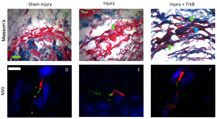
External urethral sphincter anatomy. Representative images of Masson’s stained urethral cross-sections 6 weeks after (A) sham dual injury treated with saline (Sham Injury); (B) pudendal nerve crush (PNC) and vaginal distension (VD) injury treated with saline (Injury); and (C) PNC + VD injury treated with tyrosine kinase B (TrkB) fusion-chimera (Injury + TrkB). Representative images of neuromuscular junction (NMJ) immunofluorescence 6 weeks after (D) sham injury, (E) injury, and (F) injury + TrkB. In Masson’s stained specimens (A–C), collagen was stained blue, and muscle cells were stained red. In immunofluorescence images (D–F), nerve axons are stained green, NMJs are stained red, and the muscle is stained blue. The green scale bar in panel (A) for Masson’s trichrome data is 100 µm. The white scale bar in panel (D) for immunofluorescence data is 20 µm. Green arrows indicate collagen, and the blue arrow indicates disrupted EUS.

