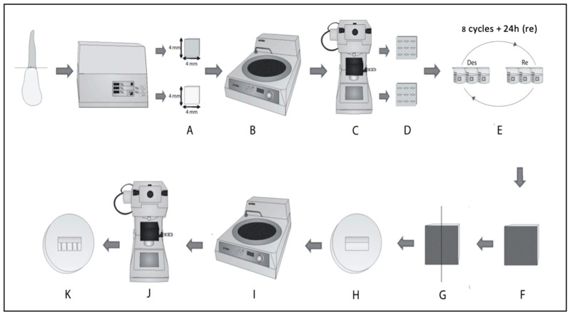
Figure 1.
Methodology used. (A) Dental blocks; (B) buccal enamel removed using an Aropol-E polisher; (C) baseline microhardness of dentin was tested; (D) dentin blocks after indentations; (E) carious lesion induction; (F) view of the demineralized surface after pH cycles; (G) specimens sectioned in half (treatment X control); (H) dentin surface to be subjected to microhardness; (I) sample polishing for final microhardness measurement; (J) microhardness analysis after 24 h as well as 7 days, 30 days, and 60 days; and (K) final orientation of the indentations in the sample [23].

