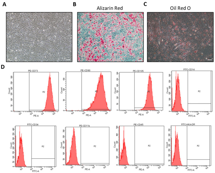
Figure 1.
Confirmation of hiMSC characteristics. (A) The fifth passage of hiMSCs cultured in vitro for 3 days was observed under a microscope. (B) Osteoblasts were identified by Alizarin Red staining, in which a red stain showed cells with calcium accumulation. (C) Adipogenesis was identified by Oil Red O staining, in which red satin showed accumulation of neutral lipid within the cells. (D) Phenotypes of hiMSCs were detected by FC. hiMSCs expressed CD73, CD90 and CD105 highly. Contrarily, the expression of CD14b, CD34, CD11b, CD45 and HLA-DR was negative in the hiMSCs. Scale bar: 200 μm.

