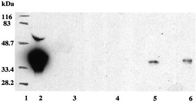
FIG. 1.
LAP expression by rBCG. Western blot analysis demonstrated the presence of LAP in cell lysates from rBCG clones 3a/LAP and 4a/LAP (lanes 5 and 6, respectively). The band corresponding to 4a/LAP runs slightly slower than that corresponding to 3a/LAP because of the addition of the HA tag. LAP was not detected in corresponding culture filtrate preparations. No reactivity was observed in cell extract or culture filtrate from control BCG transformed with the pBRD3a vector (lane 3 or 4, respectively). Lanes 1 and 2 show molecular weight markers and the antibody response to 50 ng of recombinant human LAP, respectively. LAP expression in rBCG extracts was estimated to be approximately 2.5 ng per 107 cells.

