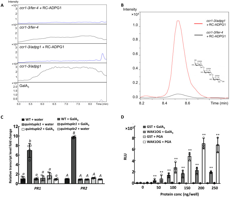
Fig. 5. Pectins released from cell walls of the ccr1 mutant are cleaved by ADPG1 to release GalA3, which binds to the WAK OG-binding domain and induces PR proteins.
(A) Extracted ion chromatograms of OGs from DP3 to DP9 in CWEs from ccr1-3/fer-4 and ccr1-3/adpg1 with and without digestion with recombinant ADPG1. (B) Selected ion monitoring for GalA3 (at m/z = 545) of LC-MS traces of oligosaccharides from CWEs from ccr1-3/fer-4 (black) and ccr1-3/adpg1 (red). CWEs were preincubated with recombinant ADPG1. (C) PR1 and PR2 transcript levels in leaves of Col-0 or wak1/2/3/4/5 quintuple mutant after injection of water (control) or GalA3 (0.05 mg/ml). (D) ELISA of the binding of GalA3 (20 μg per well) and polygalacturonic acid (20 μg per well, positive ligand control) to the WAK1 OG-binding domain, expressed in E. coli as a GST fusion protein (fig. S9B). GST serves as a negative binding protein control. Bars represent means ± SD. n = 3. Hollow dots represent individual data points in (C) and (D). Letters and asterisks indicate statistically significant differences among samples according to one-way ANOVA followed by Tukey’s HSD test, α = 0.05, in (C) and Student’s t test, ***P < 0.001, in (D).

