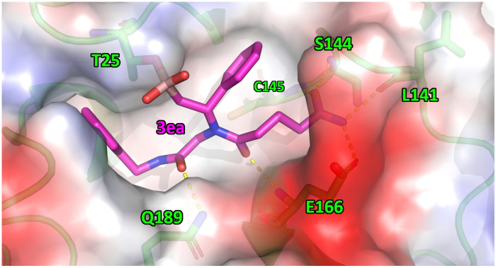
Figure 3.
Supposed binding mode of 3ea (magenta sticks) in complex with MproCoV-2. The enzyme surface is represented as a “solvent accessible surface”, in which the partial charges of the residue atoms are colored in blue or in red, accordingly with their positive or negative charge. The hydrogen bonds are represented as dashed yellow lines. Residues are numbered as was found in the X-ray structure.

