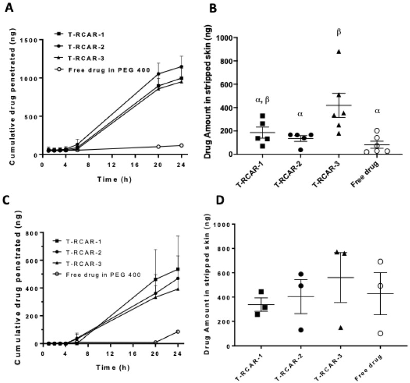
Figure 2.
Ex vivo skin drug permeation profiles of T-RCARs using 40% PEG 400 in PBS or 4% BSA in PBS as receiver media. (A) Formulations containing 4 μg drug in 0.2 mL formulation solutions were loaded to the skin facing the donor compartments in the Franz diffusion cells. Data shown are cumulative R-carvedilol permeated into the receptor compartment as a function of time up to 24 h. The receiver compartment contains PBS containing 40% v/v of PEG400. Data are presented as mean ± SE (n = 5~6). (B) R-carvedilol levels in stripped skin (epidermal and dermal layers) 24 h after loading the drugs, determined via HPLC. Data are presented as mean ± SE (n = 5~6). (C) Cumulative R-carvedilol permeated into the receptor compartment as a function of time up to 24 h. The receiver compartment contains 4% BSA in PBS. Data are presented as mean ± SE (n = 3). (D) R-carvedilol levels in stripped skin (epidermal and dermal layers) 24 h after loading the drugs, determined via HPLC. Data are presented as mean ± SE (n = 3). An ANOVA followed by a Tukey–Kramer multiple comparisons test was used to assess statistical differences at p < 0.05, and differences denoted by different Greek letters.

